

The human ribosomal protein genes: sequencing and comparative analysis of 73 genes
- PMID: 11875025
- PMCID: PMC155282
- DOI: 10.1101/gr.214202
The human ribosomal protein genes: sequencing and comparative analysis of 73 genes
Abstract
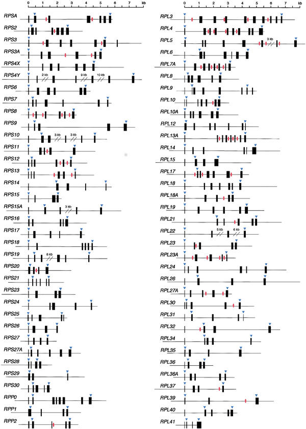
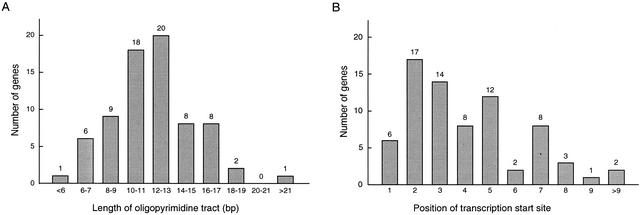
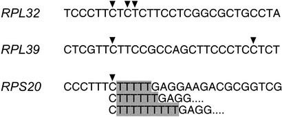
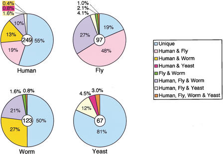
The ribosome, as a catalyst for protein synthesis, is universal and essential for all organisms. Here we describe the structure of the genes encoding human ribosomal proteins (RPs) and compare this class of genes among several eukaryotes. Using genomic and full-length cDNA sequences, we characterized 73 RP genes and found that (1) transcription starts at a C residue within a characteristic oligopyrimidine tract; (2) the promoter region is GC rich, but often has a TATA box or similar sequence element; (3) the genes are small (4.4 kb), but have as many as 5.6 exons on average; (4) the initiator ATG is in the first or second exon and is within plus minus 5 bp of the first intron boundaries in about half of cases; and (5) 5'- and 3'-UTRs are significantly smaller (42 bp and 56 bp, respectively) than the genome average. Comparison of RP genes from humans, Drosophila melanogaster, Caenorhabditis elegans, and Saccharomyces cerevisiae revealed the coding sequences to be highly conserved (63% homology on average), although gene size and the number of exons vary. The positions of the introns are also conserved among these species as follows: 44% of human introns are present at the same position in either D. melanogaster or C. elegans, suggesting RP genes are highly suitable for studying the evolution of introns.
Figures


References
-
- Amaldi F, Camacho-Vanegas O, Cardinall B, Cecconi F, Crosio C, Loreni F, Mariottini P, Pellizzoni L, Pierandrei-Amaldi P. Structure and expression of ribosomal protein genes in Xenopus laevis. Biochem Cell Biol. 1995;73:969–977. - PubMed
-
- Asakawa S, Abe I, Kudoh Y, Kishi N, Wang Y, Kubota R, Kudoh J, Kawasaki K, Minoshima S, Shimizu N. Human BAC library: Construction and rapid screening. Gene. 1997;191:69–79. - PubMed
-
- Ban N, Nissen P, Hansen J, Moore PB, Steitz TA. The complete atomic structure of the large ribosomal subunit at 2.4 Å resolution. Science. 2000;289:905–920. - PubMed
-
- Baxter LL, Moran TH, Richtsmeier JT, Troncoso J, Reeves RH. Discovery and genetic localization of Down syndrome cerebellar phenotypes using the Ts65Dn mouse. Hum Mol Genet. 2000;9:195–202. - PubMed
Publication types
MeSH terms
Substances
Associated data
- Actions
- Actions
- Actions
- Actions
- Actions
- Actions
- Actions
- Actions
- Actions
- Actions
- Actions
- Actions
- Actions
- Actions
- Actions
- Actions
- Actions
- Actions
- Actions
- Actions
- Actions
- Actions
- Actions
- Actions
- Actions
- Actions
- Actions
- Actions
- Actions
- Actions
- Actions
- Actions
- Actions
- Actions
- Actions
- Actions
- Actions
- Actions
- Actions
- Actions
- Actions
- Actions
- Actions
- Actions
- Actions
- Actions
- Actions
- Actions
- Actions
- Actions
- Actions
- Actions
- Actions
- Actions
- Actions
- Actions
- Actions
- Actions
- Actions
- Actions
- Actions
- Actions
- Actions
- Actions
- Actions
- Actions
- Actions
LinkOut - more resources
Full Text Sources
Other Literature Sources
Molecular Biology Databases
Miscellaneous