Comparative Study
. 2002 Mar 1;11(5):559-68.
doi: 10.1093/hmg/11.5.559.

Mutations in the inosine monophosphate dehydrogenase 1 gene (IMPDH1) cause the RP10 form of autosomal dominant retinitis pigmentosa

Affiliations
Comparative Study

Mutations in the inosine monophosphate dehydrogenase 1 gene (IMPDH1) cause the RP10 form of autosomal dominant retinitis pigmentosa

Sara J Bowne et al. Hum Mol Genet. .

Abstract

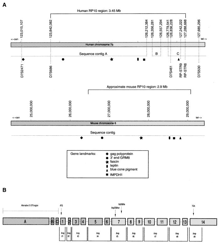
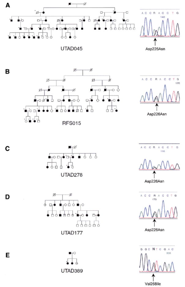
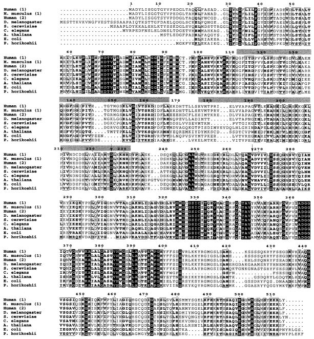
Autosomal dominant retinitis pigmentosa (adRP) is a heterogeneous set of progressive retinopathies caused by several distinct genes. One locus, the RP10 form of adRP, maps to human chromosome 7q31.1 and may account for 5-10% of adRP cases among Americans and Europeans. We identified two American families with the RP10 form of adRP by linkage mapping and used these families to reduce the linkage interval to 3.45 Mb between the flanking markers D7S686 and RP-STR8. Sequence and transcript analysis identified 54 independent genes within this region, at least 10 of which are retinal-expressed and thus candidates for the RP10 gene. A screen of retinal transcripts comparing retinas from normal mice to retinas from crx-/crx- knockout mice (with poorly differentiated photoreceptors) demonstrated a 6-fold reduction in one candidate, inosine monophosphate dehydrogenase 1 (IMPDH1; EC 1.1.1.205). Since many of the genes known to cause retinitis pigmentosa are under CRX control in photoreceptors, IMPDH1 became a high-priority candidate for mutation screening. DNA sequencing of affected individuals from the two American RP10 families revealed a GAC-->AAC transition in codon 226 substituting an asparagine for an aspartic acid in both families. The identical mutation was also found in a British RP10 family. The Asp226Asn missense mutation is present in all affected individuals tested and absent from unaffected controls. The aspartic acid at codon 226 is conserved in all IMPDH genes, in all species examined, including bacteria, suggesting that this mutation is highly deleterious. Subsequent screening of probands from 60 other adRP families revealed an additional family with this mutation, confirming its association with retinitis pigmentosa and the relatively high frequency of this mutation. Another IMPDH1 substitution, Val268Ile, was also observed in this cohort of patients but not in controls. IMPDH1 is a ubiquitously expressed enzyme, functioning as a homotetramer, which catalyzed the rate-limiting step in de novo synthesis of guanine nucleotides. As such, it plays an important role in cyclic nucleoside metabolism within photoreceptors. Several classes of drugs are known to affect IMPDH isoenzymes, including nucleotide and NAD analogs, suggesting that small-molecule therapy may be available, one day, for RP10 patients.


Figures

Figure 1
Figure 1
Representative fundus photographs, ERGs and visual fields from the affected proband of family RFS015. The proband was aged 24 years when first seen at the Retina Foundation of the Southwest in 1988. Best-corrected vision in each eye was 20/50. The lenses of both eyes showed some posterior subcapsular cataract. He had been severely night blind for many years and voluntarily stopped driving at age 17. The fundus of each eye showed typical RP with attenuated retinal vessels and bone-spicule type pigmentary abnormalities throughout the midperiphery. ERG responses were significantly reduced in amplitude. However, rod responses were clearly detectable despite being reduced by ~90% below the lower limit of normal. Cone responses to 30 Hz flicker were reduced in amplitude by 90% and borderline delayed in b-wave implicit time, as reported previously for some patients with adRP (28). This pattern of ERG loss is unusual in that there is a similar reduction in rod and cone responses and could potentially be a phenotypic marker of IMPDH1 mutations. The more usual pattern in adRP is for greater loss of rod than cone responses. Consistent with the recordable rod ERG, final dark-adapted visual thresholds were elevated 0.6 log units above the upper limit of normal. This contrasts with the 3.0 log unit elevations typically seen in patients lacking rod ERG function. Visual fields were also consistent with the ERG in showing severe constriction to the IV-4-e test target. The IV-4-e isopter was within 20 degrees.
Figure 2
Figure 2
(A) Diagram of the RP10 candidate region on human chromosome 7q31.1 and the syntenic region on mouse chromosome 6. The recombinant event in UTAD045 that defines the RP10 critical region was localized to the region between RP-STR8 and RPSTR9. The approximate location of several known genes from this chromosomal region are shown for each species. Comparison of several genes and EST clusters from mouse and human led us to believe that contig B, as well as the majority of gap sequence located on either side of contig B, is erroneously placed. (B) IMPDH1 gene structure and location of screening amplimers used in this study. Exons are labeled accorded to nomenclature initially used by Gu et al. (12). Exons 1–14 contain the coding region of all three reported IMPDH1 transcripts. Exons A–C represent alternative 5′-UTRs used only in the 4.0 and 2.7 kb IMPDH1 transcripts.
Figure 3
Figure 3
Pedigrees and electropherograms of five RP10 families with IMPDH1 mutations. All families are of American origin with the exception of UTAD278, which is a British family. (A) UTAD045, Asp226Asn; (B) RFS015, Asp226Asn; (C) UTAD278, Asp226Asn; (D) UTAD177, Asp226Asn; and (E) UTAD389, Val268Ile. Closed symbols represent affected individuals, question marks represent possibly affected individuals, and open symbols represent unaffected individuals. Tested individuals in each family are indicated by a star.
Figure 4
Figure 4
IMPDH protein sequences from a range of organisms, aligned using ClustalW and formatted using ESPript 2.0 (prodes.toulouse.inra.fr/ESPript/cgi-bin/nph-ESPript_exe.cgi). In cases where multiple isoforms have been identified, isoform numbers are in parentheses. Sequence numbering is based on the human IMPDH1 sequence. Completely conserved residues are shown as white letters on a black background; highly conserved residues are boxed. The locations of the two CBS domains are indicated by gray bars and the location of the two mutations identified in this study are shown by arrows. SwissProtID and accession numbers: human-1 (imd1_human, P20829), Mus musculus-1 (imd1_mouse, P50096), human-2 (imd2_human, P12268), Drosophila melanogaster (imdh_drome, Q07152), Saccharomyces cerevisiae (imh1_yeast, P38697), Caenorhabditis elegans (PIR T3709), Arabidopsis thaliana (imh1_arath, P47996), E. coli (imdh_ecoli, P06981) and Pyrococcus horikoshii (impd_pyrho, O58045).

References

    1. Heckenlively JR, Daiger SP. Hereditary retinal and choroidal degenerations. In: Rimoin DI, Connor JM, Pyeritz RE, editors. Principals and Practices of Medical Genetics. 4. Vol. 1. Churchill Livingston; New York: 2001. pp. 2255–2576.
    1. Sohocki MM, Daiger SP, Bowne SJ, Rodriquez JA, Northrup H, Heckenlively JR, Birch DG, Mintz-Hittner H, Ruiz RS, Lewis RA, Saperstein DA, Sullivan LS. Prevalence of mutations causing retinitis pigmentosa and other inherited retinopathies. Hum Mutat. 2001;17:42–51. - PMC - PubMed
    1. Jordan SA, Farrar GJ, Kenna P, Humphries MM, Sheils DM, Kumar-Singh R, Sharp EM, Soriano N, Ayuso C, Benitez J, et al. Localization of an autosomal dominant retinitis pigmentosa gene to chromosome 7q. Nat Genet. 1993;4:54–58. - PubMed
    1. McGuire RE, Gannon AM, Sadler-Sullivan LA, Rodriguez JA, Daiger SP. Evidence for a major gene (RP10) for autosomal dominant retinitis pigmentosa on chromosome 7q: linkage mapping in a second, unrelated family. Hum Genet. 1995;95:71–74. - PubMed
    1. McGuire RE, Jordan SA, Braden VV, Bouffard GG, Humphries P, Green ED, Daiger SP. Mapping the RP10 locus for autosomal dominant retinitis pigmentosa on 7q: refined genetic positioning and localization within a well-defined YAC contig. Genome Res. 1996;6:255–266. - PubMed

Publication types