Review
. 2002 Mar;66(1):1-20, table of contents.
doi: 10.1128/MMBR.66.1.1-20.2002.

Hydrogenases and hydrogen metabolism of cyanobacteria

Affiliations
Free PMC article
Review

Hydrogenases and hydrogen metabolism of cyanobacteria

Paula Tamagnini et al. Microbiol Mol Biol Rev. 2002 Mar.
Free PMC article

Abstract

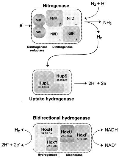
Cyanobacteria may possess several enzymes that are directly involved in dihydrogen metabolism: nitrogenase(s) catalyzing the production of hydrogen concomitantly with the reduction of dinitrogen to ammonia, an uptake hydrogenase (encoded by hupSL) catalyzing the consumption of hydrogen produced by the nitrogenase, and a bidirectional hydrogenase (encoded by hoxFUYH) which has the capacity to both take up and produce hydrogen. This review summarizes our knowledge about cyanobacterial hydrogenases, focusing on recent progress since the first molecular information was published in 1995. It presents the molecular knowledge about cyanobacterial hupSL and hoxFUYH, their corresponding gene products, and their accessory genes before finishing with an applied aspect--the use of cyanobacteria in a biological, renewable production of the future energy carrier molecular hydrogen. In addition to scientific publications, information from three cyanobacterial genomes, the unicellular Synechocystis strain PCC 6803 and the filamentous heterocystous Anabaena strain PCC 7120 and Nostoc punctiforme (PCC 73102/ATCC 29133) is included.


Figures

FIG. 1.
FIG. 1.
Enzymes directly involved in hydrogen metabolism in cyanobacteria. While the uptake hydrogenase is present in all nitrogen-fixing strains tested so far, the bidirectional enzyme is distributed among both nitrogen-fixing and non-nitrogen-fixing cyanobacteria (although it is not a universal cyanobacterial enzyme) (192). The molecular masses indicated for the uptake hydrogenase subunits are mean values calculated from the deduced amino acid sequences of Anabaena strain PCC 7120 (39), Nostoc strain PCC 73102 (150), and A. variabilis ATCC 29413 (79), while the values for the subunits of the bidirectional enzyme are based on data exclusively from A. variabilis ATCC 29413 (173).
FIG. 2.
FIG. 2.
Microphotographs of the three cyanobacteria from which genome sequence information is included in the present review. (A) Synechocystis strain PCC 6803; (B) Anabaena strain PCC 7120; (C) Nostoc punctiforme PCC 73102/ATCC 29133. Note the presence of heterocysts (arrows) in the nitrogen-fixing cultures of Anabaena strain PCC 7120 and Nostoc strain PCC 73102. Bar, 10 μm.
FIG.3.
FIG.3.
hupSL in the cyanobacteria Nostoc strain PCC 73102 (N. PCC 73102) (150), A. variabilis ATCC 29413 (79), and Anabaena strain PCC 7120 (A. PCC 7120) (39). (A) Thin vertical lines correspond to the nucleotide triplets encoding the conserved cysteine residues compared to HupSL in other organisms. Arrows show the positions of transcriptional start in Nostoc strain PCC 73102 and A. variabilis ATCC 29413. Grey boxes (a, b, c, and d) indicate the positions of putative promoter elements; a, NtcA-binding site; b, IHF-binding site; c, −10 promoter sequence; d, part of an FNR-binding site. Intergenic-sequence hairpin structures are also indicated (for details, see panel B), as well as positions and directions of putative ORFs, labeled 1 to 3, downstream of hupL. Note the presence of xisC and the subsequent rearrangement within hupL in Anabaena strain PCC 7120. (B) hupSL intergenic sequence hairpin structures. The stop codon of hupS and the start codon of hupL are boxed, and calculated ΔG values for each hairpin structure are shown below the respective structures (112).
FIG. 4.
FIG. 4.
Induction of an in vivo light-dependent hydrogen uptake in Nostoc muscorum cells during a shift from non-nitrogen-fixing to nitrogen-fixing conditions. Ammonia-grown cells were transferred to nitrogen-fixing conditions (time = 0) and analyzed with a Clark-type electrode for the appearance of an in vivo light-dependent hydrogen uptake (A) and by RT-PCR for the presence of a hupL transcript (B) after 14, 24, 41, and 61 h (17). M, marker (100-bp DNA ladder). Controls (right panel) include only a PCR (-RT; all RNA samples, only t = 0 h shown, negative control), replacement of RNA with water (H2O, negative control), and use of genomic DNA instead of RNA (DNA, positive control).
FIG. 5.
FIG. 5.
Physical map of the genes encoding the bidirectional hydrogenase (hox) and additional ORFs (labeled 1 to 7, with identical numbers indicating homologous ORFs) in the cyanobacteria A. variabilis (two strains; A. ATCC 29413 [173] and A. IAM M58 [Gen. Bank accession no. AB057405]), Anabaena strain PCC 7120 (A. PCC 7120) (http://www.kazusa.or.jpn/cyano/anabaena), Synechococcus PCC 6301 (S. PCC 6301) (29, 30), and Synechocystis PCC 6803 (S. PCC 6803) (12, 95, 144). Vertical lines correspond to the positions of triplets encoding conserved cysteine residues compared to other microorganisms (for more details, see Table 4). ▨ and formula image represent the NAD- and flavin mononucleotide-binding regions, respectively.
FIG. 6.
FIG. 6.
Visualization of a hoxH transcript from N. muscorum during a shift from non-nitrogen-fixing to nitrogen-fixing conditions (the same experiment as described in the legend to Fig. 4). Transcripts from t = 0, 14, 24, 41, and 61 h after the transfer to nitrogen-fixing conditions are shown (17). M, marker (100-bp DNA ladder).
FIG. 7.
FIG. 7.
Physical organization and conserved regions (vertical lines; for further explanations, see Table 6) of hitherto identified cyanobacterial hyp genes. The histidine-rich N terminals of hypB are indicated (formula image
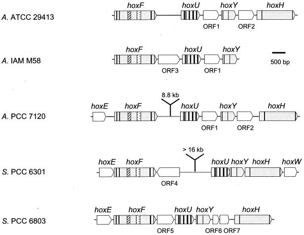
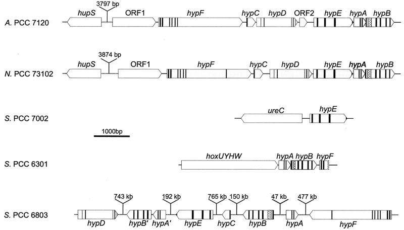
). Anabaena strain PCC 7120 (A. PCC 7120) (, ; http://www.kazusa.or.jpn/cyano/anabaena), Nostoc PCC 73102 (N. PCC 73102) (; http://www.jgi.doe.gov/JGI_microbial/html/nostoc/nostoc_homepage/html); Synechococcus strain PCC 7002 (S. PCC 7002) (171), Synechococcus strain PCC 6301 (S. PCC 6301) (29), and Synechocystis strain PCC 6803 (S. PCC 6803) (95, 144).

References

    1. Reference deleted.
    1. Reference deleted.
    1. Adams, D. G. 2000. Symbiotic interactions, p. 523-561. In B. A. Whitton and M. Potts (ed.), The ecology of cyanobacteria. Kluwer Academic Publishers, Dordrecht, The Netherlands.
    1. Akhmanova, A., F. G. Voncken, K. M. Hosea, H. Harhangi, J. T. Keltjens, H. J. op den Camp, G. D. Vogels, and J. H. Hackstein. 1999. A hydrogenosome with pyruvate formate-lyase: anaerobic chytrid fungi use an alternative route for pyruvate catabolism. Mol. Microbiol. 32:1103-1114. - PubMed
    1. Akhmanova, A., F. G. Voncken, T. van Alen, A. van Hoek, B. Boxma, G. D. Vogels, M. Veenhuis, and J. H. Hackstein. 1998. A hydrogenosome with a genome. Nature 396:527-528. - PubMed

Publication types

LinkOut - more resources