Review
. 2002 Mar;66(1):122-54; table of contents.
doi: 10.1128/MMBR.66.1.122-154.2002.

Secretory pathway of trypanosomatid parasites

Affiliations
Review

Secretory pathway of trypanosomatid parasites

Malcolm J McConville et al. Microbiol Mol Biol Rev. 2002 Mar.

Abstract

The Trypanosomatidae comprise a large group of parasitic protozoa, some of which cause important diseases in humans. These include Trypanosoma brucei (the causative agent of African sleeping sickness and nagana in cattle), Trypanosoma cruzi (the causative agent of Chagas' disease in Central and South America), and Leishmania spp. (the causative agent of visceral and [muco]cutaneous leishmaniasis throughout the tropics and subtropics). The cell surfaces of these parasites are covered in complex protein- or carbohydrate-rich coats that are required for parasite survival and infectivity in their respective insect vectors and mammalian hosts. These molecules are assembled in the secretory pathway. Recent advances in the genetic manipulation of these parasites as well as progress with the parasite genome projects has greatly advanced our understanding of processes that underlie secretory transport in trypanosomatids. This article provides an overview of the organization of the trypanosomatid secretory pathway and connections that exist with endocytic organelles and multiple lytic and storage vacuoles. A number of the molecular components that are required for vesicular transport have been identified, as have some of the sorting signals that direct proteins to the cell surface or organelles in the endosome-vacuole system. Finally, the subcellular organization of the major glycosylation pathways in these parasites is reviewed. Studies on these highly divergent eukaryotes provide important insights into the molecular processes underlying secretory transport that arose very early in eukaryotic evolution. They also reveal unusual or novel aspects of secretory transport and protein glycosylation that may be exploited in developing new antiparasite drugs.


Figures

FIG. 1.
FIG. 1.
Life cycles of trypanosomatid parasites. The insect vectors for T. brucei, T. cruzi, and Leishmania are the tsetse fly (Glossina), the reduviid bug (Triatoma), and sand flies (Phlebotomus and Lutzomyia), respectively. The mammalian stages of T. brucei exist primarily in the bloodstream. In contrast, most of the mammalian developmental stages of T. cruzi and Leishmania spp. reside within the cytoplasm of a wide range of host cells or the phagolysosome of host macrophages, respectively. Proliferative and nonproliferative (boxed) stages and the locations of the flagellum (blue) and kinetoplast (red) relative to the nucleus (grey) are indicated. T. cruzi and Leishmania amastigotes may undergo periods of proliferative and nonproliferative growth. It should be noted that most studies on the secretory functions of these parasites have been carried out on proliferative stages. Trypo, trypomastigote.
FIG. 2.
FIG. 2.
Surface coats of trypanosomatids. The surface coats of trypanosomatid parasites are dominated by GPI proteins and/or non-protein-linked GPI glycolipids. The nature of the common and species-specific modifications (N-linked glycans, O-linked glycans, and phosphoglycans) that occur in the secretory pathway of these parasites is highlighted. In Leishmania, some free GPIs are also phosphoglycosylated to form LPG.
FIG. 3.
FIG. 3.
Schematic representations of the secretory and endocytic organelles of T. brucei BF, T. cruzi epimastigotes, and Leishmania promastigotes. Most of the organelles involved in secretion and the early endocytic pathway are organized around the flagellar pocket. Significant stage- and species-specific differences exist in the organization and morphology of late-endosome and lysosomal organelles that are highlighted in these schemes. In all cases, the ER comprises the nuclear envelope (NE) and a cortical reticulum (ER) that is connected to the specialized tER proximal to the Golgi apparatus (G) and the flagellar pocket (fp). Early endosomes (EE; depicted as a complex tubule-vesicle network) are also invariably located near the flagellar pocket. In T. brucei BF, the nature of late endosomes has not been clearly defined and mature lysosomes have a predominantly perinuclear location. In Leishmania promastigotes, a population of MVBs, which may correspond to late endosomes, forms near the Golgi apparatus and fuses with the lysosome-MVT (L-MVT), which extends along the anterior-posterior axis of the cell. In T. cruzi epimastigotes, a morphologically related MVT functions as an intermediate, late-endosome (LE-MVT) compartment that transports markers from the cytostome (Cyt; a second invagination in the plasma membrane that is specialized for endocytosis) to the mature lysosomes at the aflagellate end of the cell. Acidocalcisomes (AC) constitute a second class of acidified vacuoles which contain resident proteins that are initially synthesized in the ER. fl, flagellar; mt, microtubules. See the text for references.
FIG. 4.
FIG. 4.
Secretory and endocytic organelles of L. mexicana promastigotes. Individual organelles were visualized in live L. mexicana promastigotes that expressed or were labeled with fluorescent markers, as follows. The ER is defined by a GFP chimera containing an N-terminal signal sequence and a C-terminal ER retention signal. The single Golgi apparatus is defined by a GFP chimera containing a C-terminal GRIP (TGN-binding) domain. The early endosomes are labeled with the vital dye FM 4-64 (20 min at 10°C). Endosomes and lysosomes (lysosome-MVT) are labeled with FM 4-64 (2 h at 27°C). The lysosome-MVT is labeled with a GFP chimera containing the ER glycosyltransferase dolichol-phosphate-mannose synthase, which accumulates in the lysosome-MVT in late-log- and stationary-phase promastigotes. The acidocalcisomes are labeled with the acidotrophic dye Lysotracker. Live promastigotes expressing the GFP chimeras were surface labeled with TRITC (tetramethyl rhodamine isocyanate)-concanavalin A to highlight the cell body, the flagellum, and the flagellar pocket at the anterior end of the cell (226).
FIG. 5.
FIG. 5.
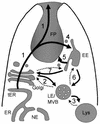
Proposed protein transport pathways in the secretory and endocytic pathways of trypanosomatids. Proteins are synthesized in the rough ER, comprising domains of the nuclear envelope (NE) and the cortical ER (ER), and then exported to the Golgi apparatus via a specialized tER. Transport from the Golgi to the flagellar pocket membrane may occur in a pleiomorphic population of vesicles and cisternal vacuoles (route 1). After delivery to the flagellar pocket, plasma membrane proteins can be directed to the cell body and/or the flagellum. Resident lysosomal proteins and ER membrane proteins destined for degradation can be transported to lysosomes via a direct internal route from the Golgi apparatus (1, 2, and 3) or after delivery to the flagellar pocket and internalization in early endosomes (1, 4, 6, and 3). Both routes may involve the internalization of membrane proteins into the lumen of MVBs as microinvaginating vesicles (2 and 6). Lysosomal resident proteins and the degradation products of lysosomes may be exocytosed via various retrograde pathways (3, 6, and 5; dotted lines) or by direct fusion with the flagellar pocket membrane. Some surface proteins are recycled through the endosomes and returned to the flagellar pocket membrane (4 and 5). Evidence for these pathways is drawn from studies on T. brucei, T. cruzi, Leishmania, and C. fasciculata (see the text for details and references). Solid lines indicate pathways that are supported by biochemical or ultrastructural studies. Dotted lines are speculative but are based on pathways that have been defined in other eukaryotes. See the text for references.
FIG. 6.
FIG. 6.
Secretory pathway modifications of proteins and GPIs in trypanosomatids. Species- and stage-specific differences in the assembly and processing of N-linked glycans, protein-linked and free GPIs, and O-linked glycans and phosphoglycosylation are highlighted. Examples of only the major glycosylation reactions are shown. The presence and location of alkylacylglycerol-ceramide remodeling reactions in T. cruzi are speculative, as is the localization of O glycosylation and phosphoglycosylation reactions in this parasite. Ara, arabinopyranose; AEP, aminoethyl-phosphonate; EP, ethanolamine phosphate; G, galactose (pyranose); Gf, galactofuranose; Glc, glucose; Gn, GlcN/GlcNAc; M, Man; Rha, rhamnose; Xyl, xylose. Note that N-glycans can also be added to the mucins and PPGs of Leishmania and T. cruzi.
FIG. 6.
FIG. 6.
Secretory pathway modifications of proteins and GPIs in trypanosomatids. Species- and stage-specific differences in the assembly and processing of N-linked glycans, protein-linked and free GPIs, and O-linked glycans and phosphoglycosylation are highlighted. Examples of only the major glycosylation reactions are shown. The presence and location of alkylacylglycerol-ceramide remodeling reactions in T. cruzi are speculative, as is the localization of O glycosylation and phosphoglycosylation reactions in this parasite. Ara, arabinopyranose; AEP, aminoethyl-phosphonate; EP, ethanolamine phosphate; G, galactose (pyranose); Gf, galactofuranose; Glc, glucose; Gn, GlcN/GlcNAc; M, Man; Rha, rhamnose; Xyl, xylose. Note that N-glycans can also be added to the mucins and PPGs of Leishmania and T. cruzi.
FIG. 7.
FIG. 7.
GPI biosynthetic pathways in T. brucei BF and Leishmania promastigotes. The T. brucei pathway has been reviewed in reference and incorporates data from recent studies (222). The L. mexicana pathway is from references and . The protein anchor pathway is not as well characterized in L. mexicana, and it is unclear whether inositol acylation is required for addition of the terminal ethanolamine phosphate (indicated by “?”). The final lipid composition of these GPIs reflects the initial incorporation of distinct PI molecular species (indicated by PIA, etc.) and/or fatty acid remodeling reactions. The fatty acid remodeling steps involve the removal of a fatty acid from the sn-1 or sn-2 position of the glycerol backbone and transfer of a new fatty acid from an acyl-CoA donor. All GPI intermediates in T. brucei BF contain a diacylglycerol lipid, and both fatty acids are remodeled. In contrast, GPI intermediates in most Leishmania spp. contain alkylacylglycerol lipids (the alkyl group is indicated by a prime) and only the sn-2 acyl group is remodeled. All the reactions shown here, with the exception of those involved in the assembly of the LPG anchor and phosphoglycan chains, occur in the ER. This figure was adapted from reference .

References

    1. Ace, T. A., S. Gokool, D. McGhie, S. Stager, and D. F. Smith. 1999. Expression of hydrophilic surface proteins in infective stages of Leishmania donovani. Mol. Biochem. Parasitol. 102:191-196. - PubMed
    1. Achleitner, G., B. Gaigg, A. Krasser, E. Kainersdorfer, S. D. Kohlwein, A. Perktold, G. Zellnig, and G. Daum. 1999. Association between the endoplasmic reticulum and mitochondria of yeast facilitates interorganelle transport of phospholipids through membrane contacts. Eur. J. Biochem. 264:545-553. - PubMed
    1. Acosta Serrano, A., S. Schenkman, N. Yoshida, A. Mehlert, J. M. Richardson, and M. A. J. Ferguson. 1995. The lipid structure of the glycosylphosphatidylinositol-anchored mucin-like sialic acid acceptors of Trypanosoma cruzi changes during parasite differentiation from epimastigotes to infective metacyclic trypomastigote forms. J. Biol. Chem. 270:27244-27253. - PubMed
    1. Acosta-Serrano, A., I. C. Almeida, L. H. Freitas-Junior, N. Yoshida, and S. Schenkman. 2001. The mucin-like glycoprotein super-family of Trypanosoma cruzi: structure and biological roles. Mol. Biochem. Parasitol. 114:143-150. - PubMed
    1. Acosta-Serrano, A., R. N. Cole, and P. T. Englund. 2000. Killing of Trypanosoma brucei by concanavalin A: structural basis of resistance in glycosylation mutants. J. Mol. Biol. 304:633-644. - PubMed

Publication types

LinkOut - more resources