

Antigen presentation by macrophages is enhanced by the uptake of necrotic, but not apoptotic, cells
- PMID: 11876743
- PMCID: PMC1906351
- DOI: 10.1046/j.1365-2249.2002.01774.x
Antigen presentation by macrophages is enhanced by the uptake of necrotic, but not apoptotic, cells
Abstract
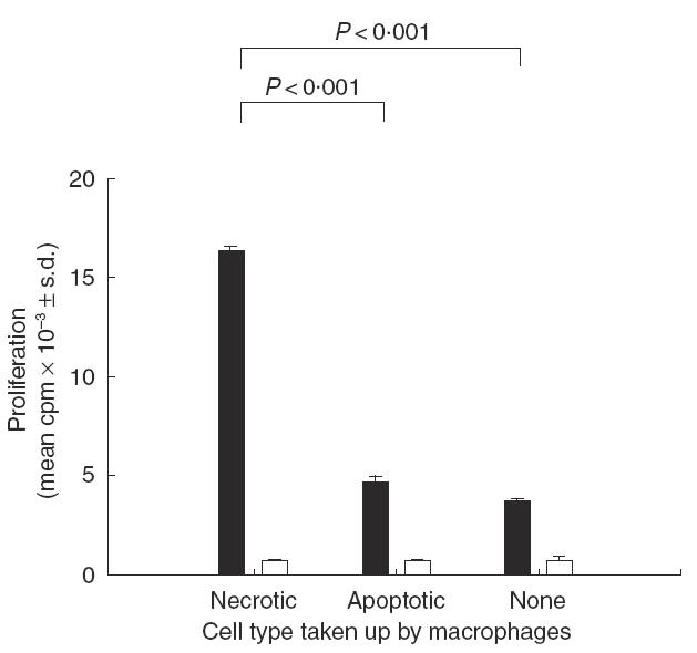
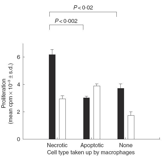
The aim of this study was to determine whether phagocytosis of necrotic or apoptotic cells affects antigen presentation by murine bone marrow-derived macrophages. After uptake of necrotic neutrophils, macrophages were able to stimulate significantly higher T cell proliferation in vitro against both the recall antigen albumin and the mitogen concanavalin A. No such effect was seen following phagocytosis of apoptotic neutrophils. Flow cytometry revealed that, within 4h of ingestion, macrophages that had taken up the necrotic cells expressed higher levels of CD40 than those that had phagocytosed apoptotic cells. Macrophage cultures pulsed with apoptotic, but not necrotic, neutrophils contained higher levels of transforming growth factor beta1, but lower concentrations of tumour necrosis factor alpha, compared to untreated controls. Our interpretation of these results is that macrophages that have taken up necrotic neutrophils co-stimulate T cells with greater efficiency due to rapid CD40 up-regulation, whereas those that have ingested apoptotic cells are not only ineffective in co-stimulation, but also secrete inhibitory cytokine.
Figures

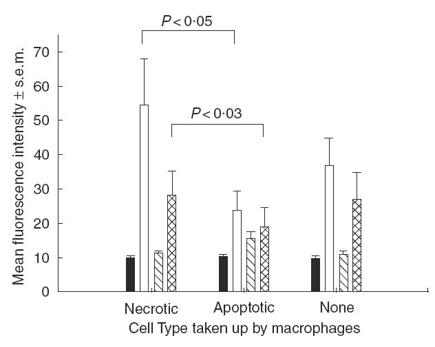
 , CD80;
 , CD86.
References
-
- Matzinger P. Tolerance, danger and the extended family. Ann Rev Immunol. 1994;12:991–1045. - PubMed
-
- Gallucci S, Lolkema M, Matzinger P. Natural adjuvants: endogenous activators of dendritic cells. Nature Med. 1999;5:1249–55. - PubMed
-
- Fearon DT, Locksley RM. The instructive role of innate immunity in the acquired immune response. Science. 1996;272:50–3. - PubMed
-
- Janeway CA. The road less travelled by. The role of innate immunity in the adaptive immune response. J Immunol. 1998;161:539–44. - PubMed
-
- Vandenbark AA, Offner H, Reshef T, et al. Specificity of T lymphocytes lines for peptides of myelin basic protein. J Immunol. 1985;135:229–33. - PubMed
Publication types
MeSH terms
Substances
LinkOut - more resources
Full Text Sources
Other Literature Sources
Research Materials
Miscellaneous
