

IgE is expressed on, but not produced by, fetal cells in the human placenta irrespective of maternal atopy
- PMID: 11876750
- PMCID: PMC1906337
- DOI: 10.1046/j.1365-2249.2002.01773.x
IgE is expressed on, but not produced by, fetal cells in the human placenta irrespective of maternal atopy
Abstract
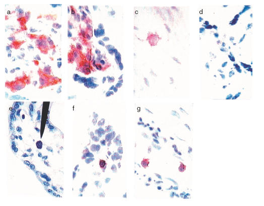
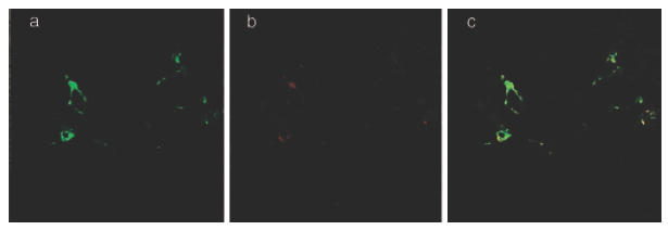
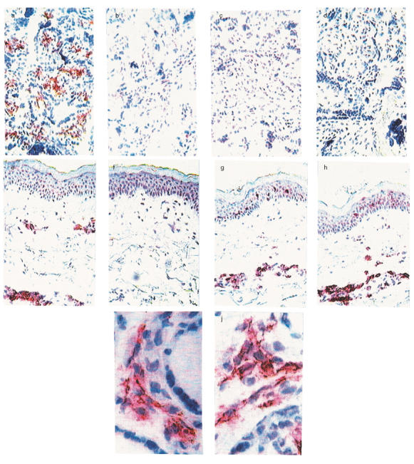
The prevalence of atopic diseases in children has increased during the last decades. Atopic symptoms usually appear early in life. This implies an early priming for atopic disease, possibly even at the fetal level. We therefore compared the presence and production of IgE in the local in utero environment during pregnancy in atopic and non-atopic women. Eighty-six women were included in the study. Fifty women were demonstrated to be atopics, based on clinical symptoms of atopic disease together with a positive Phadiatop and/or skin prick test. Placentas from these term pregnancies were obtained. Slices covering the full thickness of the placenta were cut clockwise around the umbilical cord and were analysed with immunohistochemistry. Surprisingly, numerous IgE+ cells, located primarily in the fetal villous stroma, were detected in a majority of the investigated placentas irrespective of the atopy of the mother or maternal or fetal total serum IgE levels. The placental IgE could not be demonstrated to be bound to IgE receptors, but was shown to be bound to fetal macrophages, possibly via FcgammaRI. No evidence was found for local fetal IgE production, although cells producing epsilon transcripts were occasionally detected in the decidua. We describe here the novel finding of numerous IgE+ cells in the human placenta, suggesting an hitherto unknown role for IgE in a successful pregnancy outcome, irrespective of whether or not the mother is atopic.
Figures
Similar articles
-
Presence of CD30(+) and CD30L(+) cells in human placenta and soluble CD30 levels in cord blood are independent of maternal atopy.Placenta. 2001 Apr;22(4):372-9. doi: 10.1053/plac.2000.0619. Placenta. 2001. PMID: 11286574
-
Presence of IgE cells in human placenta is independent of malaria infection or chorioamnionitis.Clin Exp Immunol. 2006 May;144(2):204-11. doi: 10.1111/j.1365-2249.2006.03055.x. Clin Exp Immunol. 2006. PMID: 16634792 Free PMC article.
-
The secretory immune system as part of the placental barrier in the second trimester of pregnancy in humans.In Vivo. 2001 Sep-Oct;15(5):429-35. In Vivo. 2001. PMID: 11695242
-
Inflammatory responses in the placenta and umbilical cord.Semin Fetal Neonatal Med. 2006 Oct;11(5):296-301. doi: 10.1016/j.siny.2006.02.011. Epub 2006 Apr 18. Semin Fetal Neonatal Med. 2006. PMID: 16621749 Review.
-
[Synthesis and modulation of IgE in the newborn infant].Allergol Immunopathol (Madr). 1998 May-Jun;26(3):87-90. Allergol Immunopathol (Madr). 1998. PMID: 9675388 Review. Spanish.
Cited by
-
Prenatal IgE as a Risk Factor for the Development of Childhood Neurodevelopmental Disorders.Front Pediatr. 2021 May 14;9:601092. doi: 10.3389/fped.2021.601092. eCollection 2021. Front Pediatr. 2021. PMID: 34055677 Free PMC article.
-
Exosomal MicroRNAs in Pregnancy Provides Insight into a Possible Cure for Cancer.Int J Mol Sci. 2020 Jul 29;21(15):5384. doi: 10.3390/ijms21155384. Int J Mol Sci. 2020. PMID: 32751127 Free PMC article.
-
IgE against food and respiratory allergens in healthy and allergic mothers and their children.Folia Microbiol (Praha). 2008;53(1):67-72. doi: 10.1007/s12223-008-0010-5. Epub 2008 May 15. Folia Microbiol (Praha). 2008. PMID: 18481221
-
Effects on neonatal immunoglobulin concentrations by infant mode of delivery in the upstate KIDS study (2008-2010).Am J Reprod Immunol. 2023 Apr;89(4):e13688. doi: 10.1111/aji.13688. Epub 2023 Feb 22. Am J Reprod Immunol. 2023. PMID: 36788284 Free PMC article.
-
Influence of atopic heredity on IL-4-, IL-12- and IFN-gamma-producing cells in in vitro activated cord blood mononuclear cells.Clin Exp Immunol. 2001 Dec;126(3):390-6. doi: 10.1046/j.1365-2249.2001.01703.x. Clin Exp Immunol. 2001. PMID: 11737052 Free PMC article.
References
-
- Cookson W. The alliance of genes and environment in asthma and allergy. Nature. 1999;402(Suppl. 6760):B5–11. - PubMed
-
- Asher MI, Weiland SK. The international study of asthma and allergies in childhood (ISAAC) ISAAC steering committee. Clin Exp Allergy. 1998;28(Suppl. 5):52–66. - PubMed
-
- Wold AE. The hygiene hypothesis revised: is the rising frequency of allergy due to changes in the intestinal flora? Allergy. 1998;53:20–5. - PubMed
-
- Alm JS, Swartz J, Lilja G, Scheynius A, Pershagen G. Atopy in children of families with an antroposophic lifestyle. Lancet. 1999;359:1485–8. - PubMed
-
- Herz U, Ahrens B, Schefold A, Joachim R, Radbruch A, Renz H. Impact of in utero Th2 immunity on T cell deviation and subsequent immediate-type hypersensitivity in the neonate. Eur J Immunol. 2000;30:714–8. 10.1002/(sici)1521-4141(200002)30:02<714::aid-immu714>3.3.co;2-3. - DOI - PubMed
Publication types
MeSH terms
Substances
LinkOut - more resources
Full Text Sources
Medical
