. 2002:3:2.
doi: 10.1186/1471-2164-3-2. Epub 2002 Feb 1.

Molecular diversity of phospholipase D in angiosperms

Affiliations

Molecular diversity of phospholipase D in angiosperms

Marek Eliás et al. BMC Genomics. 2002.

Abstract

Background: The phospholipase D (PLD) family has been identified in plants by recent molecular studies, fostered by the emerging importance of plant PLDs in stress physiology and signal transduction. However, the presence of multiple isoforms limits the power of conventional biochemical and pharmacological approaches, and calls for a wider application of genetic methodology.

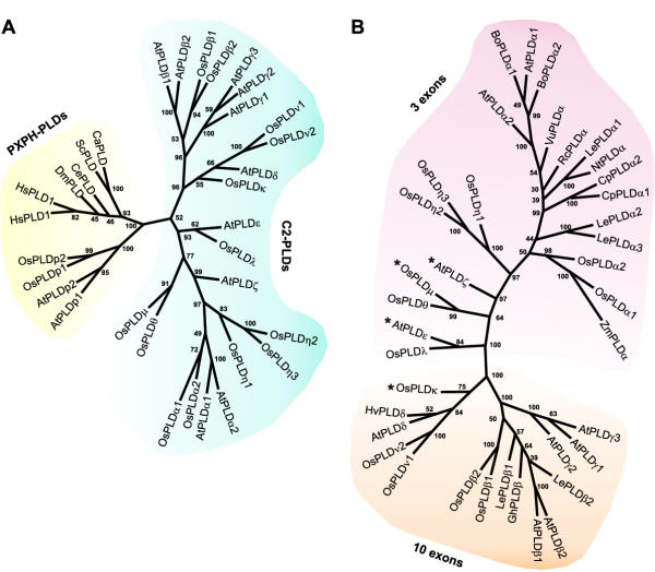
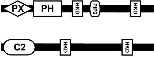
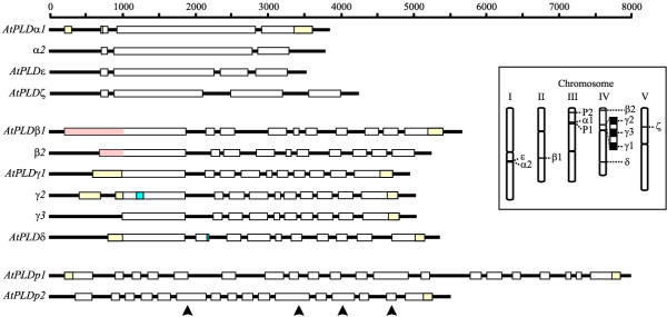
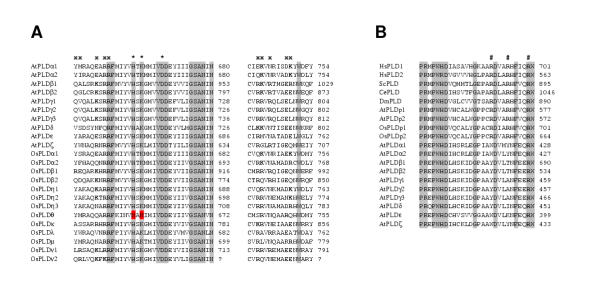
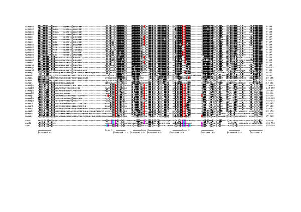
Results: Taking advantage of sequence data available in public databases, we attempted to provide a prerequisite for such an approach. We made a complete inventory of the Arabidopsis thaliana PLD family, which was found to comprise 12 distinct genes. The current nomenclature of Arabidopsis PLDs was refined and expanded to include five newly described genes. To assess the degree of plant PLD diversity beyond Arabidopsis we explored data from rice (including the genome draft by Monsanto) as well as cDNA and EST sequences from several other plants. Our analysis revealed two major PLD subfamilies in plants. The first, designated C2-PLD, is characterised by presence of the C2 domain and comprises previously known plant PLDs as well as new isoforms with possibly unusual features catalytically inactive or independent on Ca2+. The second subfamily (denoted PXPH-PLD) is novel in plants but is related to animal and fungal enzymes possessing the PX and PH domains.

Conclusions: The evolutionary dynamics, and inter-specific diversity, of plant PLDs inferred from our phylogenetic analysis, call for more plant species to be employed in PLD research. This will enable us to obtain generally valid conclusions.


Figures

Figure 1
Figure 1
Phylogenetic analysis of the PC-PLD family. A, an unrooted tree of Arabidopsis, rice and selected non-plant PLDs, constructed by the neighbor-joining method on the basis of a MACAW-generated alignment (see Materials and Methods). C2-PLD and PXPH-PLD subfamilies are indicated by yellow and blue background, respectively. Rooting the tree with a bacterial PLD (not shown) revealed that the two subfamilies are monophyletic. B, more detailed phylogeny of C2-PLDs, based on a manually edited ClustalW alignment (tree constructed as above). Note that the topology corresponds to the previous tree, with the exception of the relationships within the cluster of PLDγ. Two major monophyletic subgroups indicated by different backgrounds appear to differ in the exon-intron organisation (as inferred from data from Arabidopsis, rice, cabbage and castor bean – only cDNA sequences are available for the remaining PLDs). The number of introns in the genes marked by asterisks (*) differs secondarily from the basic plans. AtPLDε, AtPLDζ and possibly OsPLDμ have independently acquired an additional exon (see Fig. 3), and OsPLDκ has lost 4 introns corresponding to the 3rd, 7th, 8th and 9th introns of beta, gamma, delta and nu PLDs. Numbers next to the nodes are percentages of bootstrap confidence levels calculated from 500 replicates. Trees with highly congruent topology were also obtained by a maximum parsimony method. Species abbreviations: At, Arabidopsis thaliana; Bo, Brassica oleracea; Ca, Candida albicans; Ce, Caenorhabditis elegans; Cp, Craterostigma plantagineum; Dm, Drosophila melanogaster, Gh, Gossypium hirsutum; Hs, Homo sapiens; Hv, Hordeum vulgare; Le, Lycopersicon esculentum; Nt, Nicotiana tabacum; Os, Oryza sativa; Sc, Saccharomyces cerevisiae; Rc, Ricinus communis; Vu, Vigna unguiculata; Zm, Zea mays. See Additional files for accession numbers and further sequence information.
Figure 2
Figure 2
Schematic diagram of conserved motifs and domains in the two major subfamilies of PC-PLD family. PXPH-PLDs (at the top) and C2-PLDs (at the bottom) differ principally in their N-terminal regions by presence of distinct phospholipid-binding domains, i.e. the PX and PH domains in the case of the PXPH-PLD subfamily, and the C2 domain in the case of the C2-PLD subfamily. Note that the support for presence of the PX domain in plant PXPH-PLDs was slighly below the default cutoff when searched with the Search Pfam tool. The same pays true for the PX domain in AtPLDp1 identified by SMART (see Materials and Methods). HKD boxes refer to catalytic motifs forming together single catalytic site in each protein. The PIP2 box in the PXPH-PLDs relates to a PIP2-binding motif, see the text and Fig. 4B.
Figure 3
Figure 3
Exon-intron architecture and chromosomal location of PLD genes in Arabidopsis. White boxes represent coding sequences. Yellow boxes delimit un-translated regions revealed by cDNA and/or EST sequences. Blue areas in AtPLDy2 and AtPLDδ indicate potential alternative splicing (see the text). Orange regions in AtPLDβ1 and AtPLDβ2 indicate the portion of the first exon coding for the unusual N-terminal extension (details in the text). Note the three basic types of gene organisation. Black arrowheads indicate positions of introns presumably lost from the AtPLDp2 gene (compare to AtPLDp1). The inset shows locations of the PLD genes on the Arabidopsis chromosomes.
Figure 4
Figure 4
The second catalytic HKD motif and putative PIP2-binding sites in Arabidopsis and rice PLDs. A, multiple alignment of the second catalytic motif and adjacent regions harbouring alleged sites for PIP2 binding [27]. Conserved amino acid residues are indicated by shading, asterisks denote the catalytic triad. Mutated residues of the catalytic motif in OsPLDθ are on the red background. Positions of conserved residues in the postulated PIP2-binding motifs are indicated by (x), basic residues are in bold. B, multiple alignment of a box conserved among all PLDs and known to bind PIP2 in mammalian and yeast PXPH-PLDs. Three arginine residues involved in PIP2 binding [31] are absolutely conserved in all PXPH-PLDs, but are not present in C2-PLDs. Motif positions are indicated by (#), arginine residues are in bold. The numbers at the right of individual sequences in both alignments refer to the position of the last residue within the whole protein, the question mark (?) indicate that complete protein sequence is not available. Species abbreviations are the same as used in the Fig. 1. See Additional files for accession numbers and other sequence information.
Figure 5
Figure 5
Multiple alignment of C2 domains from C2-PLDs. For comparison, three characterised C2 domains (from cytosolic phospholipase A2, phospholipase Cδ and Synaptotagmin I) are included (adopted from [26]). The domain consists of eight β-strands (here indicated by lines at the bottom of the alignment) linked by loop regions. Two basic topological variants of the C2 domain have been described, resulting from a circular permutation of the β-strands (details in [26]). The Topology I is exemplified here by the C2 domain from Synaptotagmin I, whereas cPLA2 and PLCδ1 exhibit the Topology II. C2 domains from PLDs are predicted to have the Topology II [45]. The first number at each β-strand refers to the Topology II, the second number to the Topology I (the first β-strand of Synl in not shown here). Three loops containing Ca2+-coordinating ligands are indicated as Loop 1 through Loop 3. Black and grey background indicates more and less conserved positions, respectively. Residues, which bind Ca2+ by the side chains, are highlighted by a violet background. Other ligands for Ca2+ ions are provided by backbone carbonyls (the respective positions with a blue background). Only three Ca2+-binding positions (excluding backbone carbonyls) are shared by the characterised C2 domains, the first two occupied by aspartate residues, the third either by an aspartate or an asparagine residue. Other Ca2+ ligands are recruited from generally non-shared positions in distinct domains. Potential Ca2+-binding residues in the C2 domains from PLDs are shown on a red background (non-conserved residues contributing with the backbone carbonyls are not considered). No C2 domain from any PLD matches exactly any prototypic C2 domain with respect to the Ca2+-binding sites. See the text for details.

Similar articles

Cited by

References

    1. Liscovitch M, Czarny M, Fiucci G, Tang X. Phospholipase D: molecular and cell biology of a novel gene family. Biochem. 2000;345:401–415. doi: 10.1042/0264-6021:3450401. - DOI - PMC - PubMed
    1. Wang X. Multiple forms of phospholipase D in plants: the gene family, catalytic and regulatory properties, and cellular functions. Prog Lipid Res. 2000;39:109–149. doi: 10.1016/S0163-7827(00)00002-3. - DOI - PubMed
    1. Hanahan DJ, Chaikoff IL. A new phospholipide-splitting enzyme specific for the ester linkage between the nitrogenous base and the phosphoric acid grouping. J Biol Chem. 1947;169:699–705. - PubMed
    1. Wang X, Xu L, Zheng L. Cloning and expression of phosphatidylcholine-hydrolyzing phospholipase D from Ricinus communis L. J Biol Chem. 1994;269:20312–20317. - PubMed
    1. den Hartog M, Musgrave A, Munnik T. Nod factor-induced phosphatidic acid and diacylglycerol pyrophosphate formation: a role for phospholipase C and D in root hair deformation. Plant J. 2001;25:55–65. doi: 10.1046/j.1365-313X.2001.00931.x. - DOI - PubMed