. 2002 Mar;135(5):1133-43.
doi: 10.1038/sj.bjp.0704551.

Characterization of an apamin-sensitive small-conductance Ca(2+)-activated K(+) channel in porcine coronary artery endothelium: relevance to EDHF

Affiliations

Characterization of an apamin-sensitive small-conductance Ca(2+)-activated K(+) channel in porcine coronary artery endothelium: relevance to EDHF

M P Burnham et al. Br J Pharmacol. 2002 Mar.

Abstract

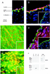
1. The apamin-sensitive small-conductance Ca(2+)-activated K(+) channel (SK(Ca)) was characterized in porcine coronary arteries. 2. In intact arteries, 100 nM substance P and 600 microM 1-ethyl-2-benzimidazolinone (1-EBIO) produced endothelial cell hyperpolarizations (27.8 +/- 0.8 mV and 24.1 +/- 1.0 mV, respectively). Charybdotoxin (100 nM) abolished the 1-EBIO response but substance P continued to induce a hyperpolarization (25.8 +/- 0.3 mV). 3. In freshly-isolated endothelial cells, outside-out patch recordings revealed a unitary K(+) conductance of 6.8 +/- 0.04 pS. The open-probability was increased by Ca(2+) and reduced by apamin (100 nM). Substance P activated an outward current under whole-cell perforated-patch conditions and a component of this current (38%) was inhibited by apamin. A second conductance of 2.7 +/- 0.03 pS inhibited by d-tubocurarine was observed infrequently. 4. Messenger RNA encoding the SK2 and SK3, but not the SK1, subunits of SK(Ca) was detected by RT - PCR in samples of endothelium. Western blotting indicated that SK3 protein was abundant in samples of endothelium compared to whole arteries. SK2 protein was present in whole artery nuclear fractions. 5. Immunofluorescent labelling confirmed that SK3 was highly expressed at the plasmalemma of endothelial cells and was not expressed in smooth muscle. SK2 was restricted to the peri-nuclear regions of both endothelial and smooth muscle cells. 6. In conclusion, the porcine coronary artery endothelium expresses an apamin-sensitive SK(Ca) containing the SK3 subunit. These channels are likely to confer all or part of the apamin-sensitive component of the endothelium-derived hyperpolarizing factor (EDHF) response.


Figures
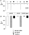
Figure 1
Figure 1
Determination using charybdotoxin and apamin of the Ca2+-activated K+ channels activated by substance P (SP) and 1-EBIO. Membrane potential responses in porcine coronary artery endothelium to transient exposure to 100 nM substance P and 600 μM 1-EBIO were recorded using sharp micro-electrodes. The addition of 100 nM charybdotoxin (ChTX) and 100 nM apamin (apa) to the superfusing bath solution is indicated. A representative trace (A) and the data derived from four such experiments (B: columns represent the mean membrane potential (Vm) and s.e.mean before and after exposure to each hyperpolarizing agent in the absence or presence of the inhibitors as indicated) is shown. Levcromakalim (LK, 10 μM) was included as an internal control.
Figure 2
Figure 2
Outside-out patch recordings from freshly-dissociated endothelial cells. Two types of small conductance channel were observed in single-channel recordings obtained with 250 nM free Ca2+ pipette solution in the presence of 100 nM charybdotoxin (physiological K+ gradient). Representative traces for each type of channel, recorded at the indicated membrane potentials are shown in (A) and (B) in which ‘c' represents the closed state. Unitary conductances were determined by variance analysis (with the assumption of a single channel) of activity recorded at −20, 0, 20 and 40 mV. Data were fitted to give slope conductances of 6.8 and 2.7 pS for each channel (C and D).
Figure 3
Figure 3
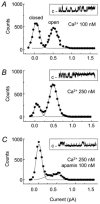
Effect of Ca2+ and apamin on the 6.8 pS channel activity. Data obtained in the outside-out patch configuration under a quasi-physiological K+ gradient (in the absence of charybdotoxin). Distribution of unitary conductance amplitude recorded at 0 mV together with representative traces (‘c' indicates the closed state): (A), with 100 nM Ca2+ pipette solution; (B), with 250 nM Ca2+ pipette solution; (C), with 250 nM Ca2+ pipette solution after the application of 100 nM apamin.
Figure 4
Figure 4
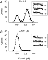
Effect of d-tubocurarine on the 2.7 pS channel activity. Data obtained at 0 mV from outside-out patches under a quasi-physiological K+ gradient with 500 nM Ca2+ pipette solution. Unitary conductance amplitude distributions together with representative traces (‘c' indicates the closed state): (A), control; (B), with 1 μM d-tubocurarine (d-TC) in the bathing solution.
Figure 5
Figure 5
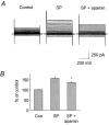
Effect of substance P on K+ currents recorded using the perforated-patch configuration. K+ currents were recorded under control conditions and during the steady state phase of current activation by 100 nM substance P in the presence and absence of 100 nM apamin. (A), superimposed traces represent K+ currents elicited by 10 mV step pulses ranging from −100 mV to +90 mV from a holding potential of −60 mV with 500 nM free Ca2+ pipette solution. (B), bar graph representing steady-state activation of K+ currents by substance P (SP) and a partial but significant block of substance P-activated current by 100 nM apamin at 90 mV (n=6).
Figure 6
Figure 6
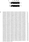
Detection of SKCa subunit mRNA in porcine coronary artery endothelium. (A), RT – PCR analysis of GAPDH (G), SK1, SK2 and SK3 expression in samples of endothelium (Endo.) and positive control pig brain prepared with (+) and without (−) reverse transcription. Size markers in base pairs are indicated. In endothelium cDNA, GAPDH (n=5/5), SK3 (n=5/5) and SK2 (n=3/5) were detected but not SK1 (n=5). (B), the complete SK3 sequence from porcine coronary artery endothelium was determined by RT – PCR and 5′ and 3′ RACE. Start and stop codons are underlined.
Figure 7
Figure 7
Localization of SK3 and SK2 protein in porcine coronary arteries by immunofluorescence labelling and Western blotting. (A), positive identification by von Willebrand's factor immunoreactivity (red) of the endothelium (Endo.) in a cross-section. Positions of the lumen, internal elastic lamina (IEL, green), smooth muscle (S. Muscle) and nuclei (blue) are shown. (B), cross-section labelled for SK3 (red) and nuclei (blue), with IEL (green) visible (inset, primary antibody pre-incubated with control peptide). (C), arteries opened longitudinally allowing the endothelium to be viewed en-face were labelled for SK3 (green) and nuclei (red) (inset, primary antibody omitted). (D), cross-section labelled for SK2 (red) and nuclei (blue), with IEL (green) shown (inset, primary antibody pre-incubated with control peptide). (E), endothelium viewed en-face and labelled for SK2 (green) and nuclei (red) (inset, primary antibody omitted). Areas where green SK2 label and red nuclei overlap appear yellow or orange. All scale bars are 25 μm. (F), Western blot analysis of SK3 and SK2 protein expression. Whole artery samples were separated into post-nuclear (P) and nuclear (N) fractions. Additional samples were prepared from endothelium (Endo.) only. Primary antibodies were used with (+) and without (−) control peptide pre-incubation. Molecular weight markers are indicated (kDa). 10 μg protein loadings.

Similar articles

Cited by

References

    1. BARFOD E.T., MOORE A.L., LIDOFSKY S.D. Cloning and functional expression of a liver isoform of the small conductance Ca2+-activated K+ channel SK3. Am. J. Physiol. 2001;280:C836–C842. - PubMed
    1. BRADFORD M.M. A rapid and sensitive method for the quantification of microgram quantities of protein utilising the principle of protein-dye binding. Anal. Biochem. 1976;72:248–254. - PubMed
    1. BURNHAM M.P., RICHARDS G.R., EDWARDS G., WESTON A.H.SK3 Ca2+-activated K+ channels are located on endothelia that generate apamin-sensitive EDHF responses Proc. Western Pharmacol. Soc. 2001(In press)
    1. BYCHKOV R., GOLLASCH M., RIED C., LUFT F.C., HALLER H. Effects of pinacidil on K+ channels in human coronary artery vascular smooth muscle cells. Am. J. Physiol. 1997;273:C161–C171. - PubMed
    1. CAO Y.-J., DREIXLER J.C., ROIZEN J.D., ROBERTS M.T., HOUAMED K.M. Modulation of recombinant small-conductance Ca2+-activated K+ channels by the muscle relaxant chlorzoxazone and structurally related compounds. J. Pharmacol. Exp. Ther. 2001;296:683–689. - PubMed

Publication types

MeSH terms

Substances