. 2002 Mar 1;16(5):560-70.
doi: 10.1101/gad.970602.

Involvement of the cohesin protein, Smc1, in Atm-dependent and independent responses to DNA damage

Affiliations

Involvement of the cohesin protein, Smc1, in Atm-dependent and independent responses to DNA damage

Seong-Tae Kim et al. Genes Dev. .

Abstract

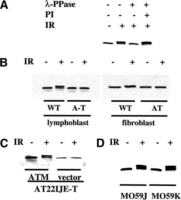
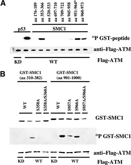
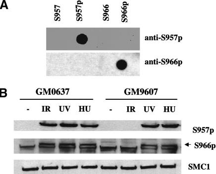
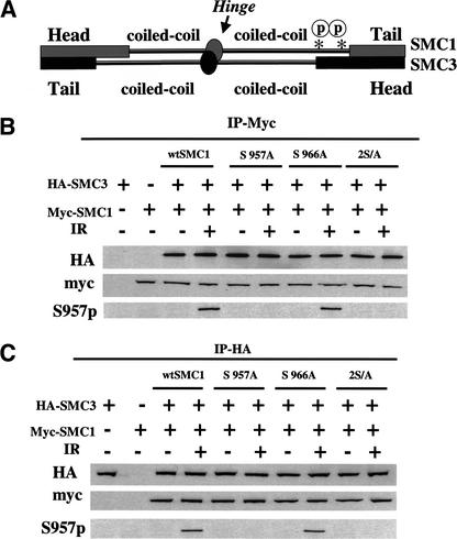
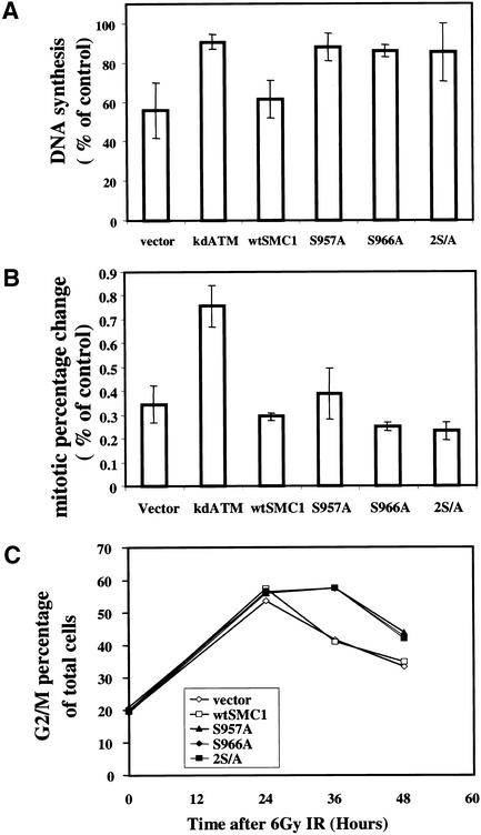
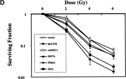
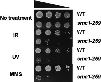
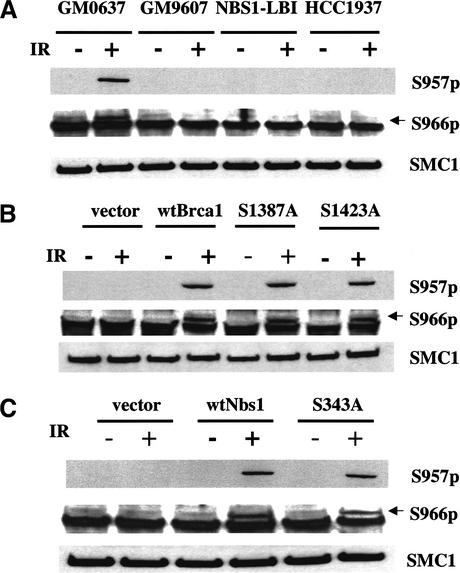
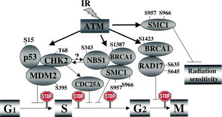
Structural maintenance of chromosomes (SMC) proteins play important roles in sister chromatid cohesion, chromosome condensation, sex-chromosome dosage compensation, and DNA recombination and repair. Protein complexes containing heterodimers of the Smc1 and Smc3 proteins have been implicated specifically in both sister chromatid cohesion and DNA recombination. Here, we show that the protein kinase, Atm, which belongs to a family of phosphatidylinositol 3-kinases that regulate cell cycle checkpoints and DNA recombination and repair, phosphorylates Smc1 protein after ionizing irradiation. Atm phosphorylates Smc1 on serines 957 and 966 in vitro and in vivo, and expression of an Smc1 protein mutated at these phosphorylation sites abrogates the ionizing irradiation-induced S phase cell cycle checkpoint. Optimal phosphorylation of these sites in Smc1 after ionizing irradiation also requires the presence of the Atm substrates Nbs1 and Brca1. These same sites in Smc1 are phosphorylated after treatment with UV irradiation or hydroxyurea in an Atm-independent manner, thus demonstrating that another kinase must be involved in responses to these cellular stresses. Yeast containing hypomorphic mutations in SMC1 and human cells overexpressing Smc1 mutated at both of these phosphorylation sites exhibit decreased survival following ionizing irradiation. These results demonstrate that Smc1 participates in cellular responses to DNA damage and link Smc1 to the Atm signal transduction pathway.


Figures

Figure 1
Figure 1
Smc1 is phosphorylated in an Atm-dependent manner after ionizing irradiation. (A) Ionizing irradiation-induced Smc1 modification is caused by serine/threonine phosphorylation. Smc1 immunoprecipitates from unirradiated or irradiated (10 Gy) normal lymphoblast cells (GM0536) were incubated with either lambda phosphatase (λ-PPase) alone or with λ-PPase and phosphatase inhibitor (PI). (IR) Ionizing irradiation. (BD) Immunoblot analyses for Smc1 protein 1 h after exposure to 0 (−) or 10 (+) Gy of ionizing irradiation in lymphoblasts [GM0536 (WT) and GM01526 (A-T)] or SV40-transformed fibroblasts [GM0637 (WT) and GM09607 (AT)] (B); A-T fibroblasts (AT22IJE-T) transfected with vector alone or with a full-length ATM cDNA (ATM) (C); or DNA-PK-deficient cells (M059J) and DNA-PK-proficient cells (M059K) (D).
Figure 2
Figure 2
Serine 957 and Serine 966 are the two major residues of Smc1 that are phosphorylated by Atm in vitro. (A) Immunoprecipitated wild-type (WT) Flag-tagged Atm or kinase-dead (KD) Flag-tagged Atm were incubated with recombinant proteins consisting of fusions between GST and peptides derived from various regions of human Smc1. The positions of the amino acids corresponding to each peptide are indicated at the top. A p53 peptide (amino acids 9–21) was used as a positive control. (*) Serine 951 was changed to alanine. (B) Atm phosphorylation of large fragments of Smc1 and mutation of relevant serines.
Figure 3
Figure 3
Atm-dependent and -independent phosphorylation of Smc1 on Ser 957 and Ser 966 in vivo. (A) Immunoblots probing 50 ng of phosphorylated or unphosphorylated synthetic peptides using rabbit polyclonal antibodies raised against peptides containing Ser 957-p or Ser 966-p. (B) Immunoblot analyses using the two phosphoserine-specific antibodies (top and middle) or an anti-SMC1 antibody (K1; bottom) on SMC1 protein from normal (WT; GM0637) or A-T (GM9607) fibroblasts treated with ionizing irradiation (IR; 10 Gy, 2 h), ultraviolet light (UV; 50 Jm−2, 2 h) or hydroxyurea (HU; 1 mM, 24 h). The arrow (middle) points to the band specifically recognized by the anti-phosphoserine 966 antibody (note a nonspecific band runs just below the S966-p band).
Figure 4
Figure 4
Phosphorylation of Smc1 does not inhibit the binding of Smc1 to Smc3. (A) Schematic representation of the domains of Smc1 bound to Smc3 illustrating the approximate location of the Smc1 Ser 957 and Ser 966 sites with the asterisk. (B) Two hours after exposure to 0 (−) or 10 (+) Gy of ionizing irradiation (IR), Myc-tagged proteins were immunoprecipitated from 293T cells that had been cotransfected with Myc-tagged SMC1 (wild type, S957A, S966A, 2S/A) and HA-tagged SMC3. The proteins were immunoblotted either with anti-Myc antibody, anti-HA antibody, or anti-S957-p antibody. (C) Two hours after exposure to 0 (−) or 10 (+) Gy of ionizing irradiation, HA-tagged proteins were immunoprecipitated from 293T cells that had been cotransfected with Myc-tagged SMC1 (wild type, S957A, S966A, 2S/A) and HA-tagged SMC3. The proteins were immunoblotted either with anti-HA antibody, anti-Myc antibody, or anti-S957-p antibody.
Figure 5
Figure 5
Mutation of the Atm phosphorylation sites on Smc1 results in an S phase checkpoint defect and radiosensitivity. (A) 293T cells transiently transfected with empty vector or wild-type (WT) or mutant (S957A, S966A, or 2S/A) of SMC1 were assessed for inhibition of DNA synthesis 30 min after exposure to 10 Gy of ionizing irradiation. (Error bars) Average of at least triplicate samples. (B) Ionizing irradiation-induced G2/M checkpoint: The mitotic percentage change of 293T cells transfected with empty vector, wild-type (WT), or mutant (S957A, S966A, or 2S/A) SMC1 or kinase-dead ATM (kdATM) 90 min after 6 Gy of ionizing irradiation is shown. Mitotic cell percentage was determined by anti-phosphohistone H3 staining followed by flow cytometric analysis. (Error bars) Average of at least triplicate samples. (C) Ionizing irradiation induced G2 accumulation: 293T cells transfected with empty vector, wild-type (WT), or mutant (S957A, S966A or 2S/A) SMC1 were irradiated at 6 Gy, and cell cycle distribution after ionizing irradiation in the indicated time points was assessed by propidium iodide staining. G2/M percentage of total cells was shown. All results are representative samples of three different experiments. (D) Expression of Smc1 phosphorylation-site mutants increases radiosensitivity. HeLa cells expressing either empty vector, wild-type (WT), or mutant (S957A, S966A, or 2S/A) constructs of SMC1 or kinase-dead ATM (kdATM) were exposed to 0–6 Gy of ionizing irradiation and incubated for 1 week prior to fixation, staining, and assessment of colony formation. The clonogenic survival assays were performed in triplicate. Error bars indicate S.E.
Figure 5
Figure 5
Mutation of the Atm phosphorylation sites on Smc1 results in an S phase checkpoint defect and radiosensitivity. (A) 293T cells transiently transfected with empty vector or wild-type (WT) or mutant (S957A, S966A, or 2S/A) of SMC1 were assessed for inhibition of DNA synthesis 30 min after exposure to 10 Gy of ionizing irradiation. (Error bars) Average of at least triplicate samples. (B) Ionizing irradiation-induced G2/M checkpoint: The mitotic percentage change of 293T cells transfected with empty vector, wild-type (WT), or mutant (S957A, S966A, or 2S/A) SMC1 or kinase-dead ATM (kdATM) 90 min after 6 Gy of ionizing irradiation is shown. Mitotic cell percentage was determined by anti-phosphohistone H3 staining followed by flow cytometric analysis. (Error bars) Average of at least triplicate samples. (C) Ionizing irradiation induced G2 accumulation: 293T cells transfected with empty vector, wild-type (WT), or mutant (S957A, S966A or 2S/A) SMC1 were irradiated at 6 Gy, and cell cycle distribution after ionizing irradiation in the indicated time points was assessed by propidium iodide staining. G2/M percentage of total cells was shown. All results are representative samples of three different experiments. (D) Expression of Smc1 phosphorylation-site mutants increases radiosensitivity. HeLa cells expressing either empty vector, wild-type (WT), or mutant (S957A, S966A, or 2S/A) constructs of SMC1 or kinase-dead ATM (kdATM) were exposed to 0–6 Gy of ionizing irradiation and incubated for 1 week prior to fixation, staining, and assessment of colony formation. The clonogenic survival assays were performed in triplicate. Error bars indicate S.E.
Figure 6
Figure 6
Smc1 protein is important for DNA damage responses in yeast. Wild-type or smc1259 mutant yeast were serially diluted by tenfold amounts and plated on YPD media at 30°C with or without 0.02% of methymethane sulfonate (MMS). Cells on plates were treated with ionizing irradiation (200 Gy) or ultraviolet light (UV; 50 Jm−2).
Figure 7
Figure 7
Brca1 and Nbs1 are required for optimal ionizing irradiation-induced Smc1 phosphorylation. (A) Smc1 phosphorylation on Ser 957 and Ser 966 is defective in cells with mutated Atm, Nbs1, or Brca1 genes: Smc1 phosphorylation was assessed by immunoblot analyses using the two phosphoserine-specific antibodies (top and middle) or an anti-Smc1 antibody (K1; bottom panel) in normal (GM0637), Atm-deficient (GM9607), Nbs1-deficient (NBS1-LBI), and Brca1-deficient (HCC1937) cell lines. (Arrows) Specific S966-p band. (B) Introduction of Brca1 into HCC1937 cells restores effective ionizing irradiation-induced Smc1 phosphorylation. Smc1 phosphorylation was assessed in HCC1937 cells that had been transiently transfected with wild-type BRCA1 (wtBrca1) or mutant [serine 1387 to alanine substitution (S1387A) or serine 1423 to alanine substitution (S1423A)] Brca1 constructs. (C) Introduction of NBS1 into NBS cells restores effective Smc1 phosphorylation. Smc1 phosphorylation was assessed in NBS cells that had been stably transfected with wild-type NBS1 (wtNbs1) or mutant [serine 343 to alanine substitution (S343A)].
Figure 8
Figure 8
Schematic representation of direct targets of ATM that affect cell cycle perturbations and modulation of sensitivity to ionizing irradiation. Following ionizing irradiation, the Atm protein kinase is activated and then phosphorylates target proteins. The exact serines (S) or threonine (T) known to be phosphorylated by Atm are listed by number for the various proteins. Chk2, p53, and Mdm2 participate in the G1 arrest, phosphorylation of Nbs1, Brca1, Smc1, and Chk2 have been implicated in controlling the S phase arrest, and phosphorylation of Brca1 and Rad17 are reported to be involved in the G2 arrest. Although phosphorylation of Chk2 has been reported to be dependent on Nbs1 (Buscemi et al. 2001), the exact relationship of the Chk2/Cdc25A axis to Nbs1/Brca1/Smc1 in controlling the S phase checkpoint remains to be clarified. On the basis of the data shown here, phosphorylation of serines 957 and 966 in Smc1 are included as targets involved in the ionizing irradiation-induced S phase arrest and are also implicated in modulating radiation sensitivity.

References

    1. Ahn J-Y, Schwarz JK, Piwnica-Worms H, Canman CE. Threonine 68 phosphorylation by ATM is required for efficient activation of Chk2 in response to ionizing radiation. Cancer Res. 2000;60:5934–5936. - PubMed
    1. Banin S, Moyal L, Shieh S-Y, Taya Y, Anderson CW, Chessa L, Smorodinsky NI, Prives C, Reiss Y, Shiloh Y, et al. Enhanced phosphorylation of p53 by ATM in response to DNA damage. Science. 1998;281:1674–1677. - PubMed
    1. Bao S, Tibbetts RS, Brumbaug KM, Fang Y, Richardson DA, Ali A, Chen SM, Abraham RT, Wang X-F. ATR/ATM-mediated phosphorylation of human Rad17 is required for genotoxic stress responses. Nature. 2001;411:969–974. - PubMed
    1. Buscemi G, Savio, Zannini L, Micciche F, Masnada D, Nakanishi M, Tauchi H, Komatsu K, Mizutani S, Khanna K, et al. Chk2 activation dependence on Nbs1 after DNA damage. Mol Cell Biol. 2001;21:5214–5222. - PMC - PubMed
    1. Canman CE, Lim D-S, Cimprich KA, Taya Y, Tamai K, Sakaguchi K, Appella E, Kastan MB, Siliciano JD. Activation of the ATM kinase by ionizing radiation and phosphorylation of p53. Science. 1998;281:1677–1679. - PubMed

Publication types

MeSH terms