

In vivo binding of active heat shock transcription factor 1 to human chromosome 9 heterochromatin during stress
- PMID: 11877455
- PMCID: PMC2173303
- DOI: 10.1083/jcb.200109018
In vivo binding of active heat shock transcription factor 1 to human chromosome 9 heterochromatin during stress
Abstract
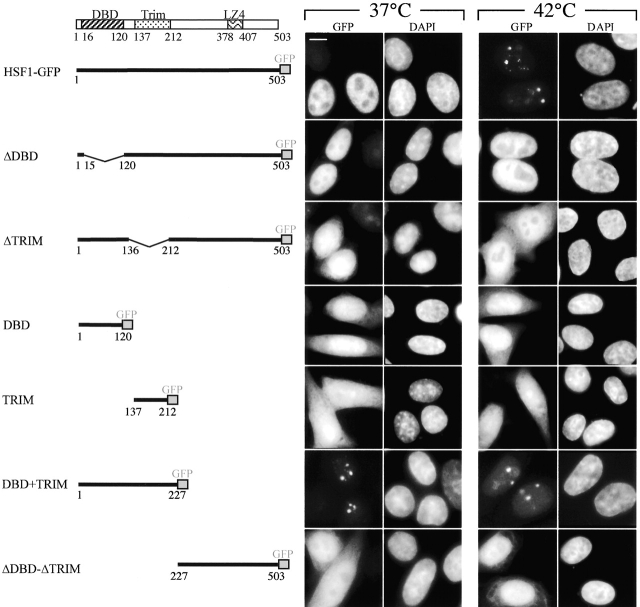
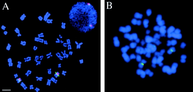
Activation of the mammalian heat shock transcription factor (HSF)1 by stress is a multistep process resulting in the transcription of heat shock genes. Coincident with these events is the rapid and reversible redistribution of HSF1 to discrete nuclear structures termed HSF1 granules, whose function is still unknown. Key features are that the number of granules correlates with cell ploidy, suggesting the existence of a chromosomal target. Here we show that in humans, HSF1 granules localize to the 9q11-q12 heterochromatic region. Within this locus, HSF1 binds through direct DNA-protein interaction with a nucleosome-containing subclass of satellite III repeats. HSF1 granule formation only requires the DNA binding competence and the trimerization of the factor. This is the first example of a transcriptional activator that accumulates transiently and reversibly on a chromosome-specific heterochromatic locus.
Figures


Similar articles
-
Stress-induced transcription of satellite III repeats.J Cell Biol. 2004 Jan 5;164(1):25-33. doi: 10.1083/jcb.200306104. Epub 2003 Dec 29. J Cell Biol. 2004. PMID: 14699086 Free PMC article.
-
Nuclear stress granules: the awakening of a sleeping beauty?J Cell Biol. 2004 Jan 5;164(1):15-7. doi: 10.1083/jcb.200311102. J Cell Biol. 2004. PMID: 14709538 Free PMC article. Review.
-
Transcriptional activation of a constitutive heterochromatic domain of the human genome in response to heat shock.Mol Biol Cell. 2004 Feb;15(2):543-51. doi: 10.1091/mbc.e03-07-0487. Epub 2003 Nov 14. Mol Biol Cell. 2004. PMID: 14617804 Free PMC article.
-
Uncoupling Stress-Inducible Phosphorylation of Heat Shock Factor 1 from Its Activation.Mol Cell Biol. 2015 Jul;35(14):2530-40. doi: 10.1128/MCB.00816-14. Epub 2015 May 11. Mol Cell Biol. 2015. PMID: 25963659 Free PMC article.
-
Polymorphic variants on chromosomes probably play a significant role in infertility.Reprod Biomed Online. 2005 Dec;11(6):726-32. doi: 10.1016/s1472-6483(10)61691-4. Reprod Biomed Online. 2005. PMID: 16417737 Review.
Cited by
-
DAXX interacts with heat shock factor 1 during stress activation and enhances its transcriptional activity.Proc Natl Acad Sci U S A. 2004 Mar 23;101(12):4100-5. doi: 10.1073/pnas.0304768101. Epub 2004 Mar 11. Proc Natl Acad Sci U S A. 2004. PMID: 15016915 Free PMC article.
-
Anaphase-promoting complex/cyclosome participates in the acute response to protein-damaging stress.Mol Cell Biol. 2010 Dec;30(24):5608-20. doi: 10.1128/MCB.01506-09. Epub 2010 Oct 11. Mol Cell Biol. 2010. PMID: 20937767 Free PMC article.
-
Reversible phase separation of HSF1 is required for an acute transcriptional response during heat shock.Nat Cell Biol. 2022 Mar;24(3):340-352. doi: 10.1038/s41556-022-00846-7. Epub 2022 Mar 7. Nat Cell Biol. 2022. PMID: 35256776
-
The role of nuclear bodies in gene expression and disease.Biology (Basel). 2013 Jul 9;2(3):976-1033. doi: 10.3390/biology2030976. Biology (Basel). 2013. PMID: 24040563 Free PMC article.
-
Mechanistic Insights Into Oxidative Response of Heat Shock Factor 1 Condensates.JACS Au. 2025 Jan 30;5(2):606-617. doi: 10.1021/jacsau.4c00578. eCollection 2025 Feb 24. JACS Au. 2025. PMID: 40017748 Free PMC article.
References
-
- Archidiacono, N., R. Antonacci, R. Marzella, P. Finelli, A. Lonoce, and M. Rocchi. 1995. Comparative mapping of human alphoid sequences in great apes, using fluorescent in situ hybridization. Genomics. 25:477–484. - PubMed
-
- Bobrow, M., K. Madan, and P.L. Pearson. 1972. Staining of some specific regions of human chromosomes, particularly the secondary constriction of No. 9. Nature. 238:122–124. - PubMed
-
- Boue, A., J. Boue, and A. Gropp. 1985. Cytogenetics of pregnancy wastage. Adv. Hum. Genet. 14:1–57. - PubMed
-
- Brown, K.E., S.S. Guest, S.T. Smale, K. Hahm, M. Merkenschlager, and A.G. Fisher. 1997. Association of transcriptionally silent genes with Ikaros complexes at centromeric heterochromatin. Cell. 91:845–854. - PubMed
Publication types
MeSH terms
Substances
Grants and funding
LinkOut - more resources
Full Text Sources
Other Literature Sources
