. 2002 Mar 4;156(5):829-41.
doi: 10.1083/jcb.200107041. Epub 2002 Mar 4.

The role of cell cycle-regulated expression in the localization of spatial landmark proteins in yeast

Affiliations

The role of cell cycle-regulated expression in the localization of spatial landmark proteins in yeast

Laura R Schenkman et al. J Cell Biol. .

Abstract

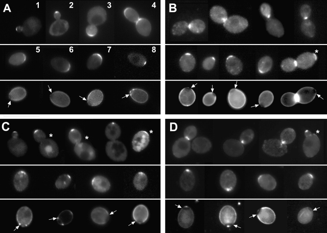
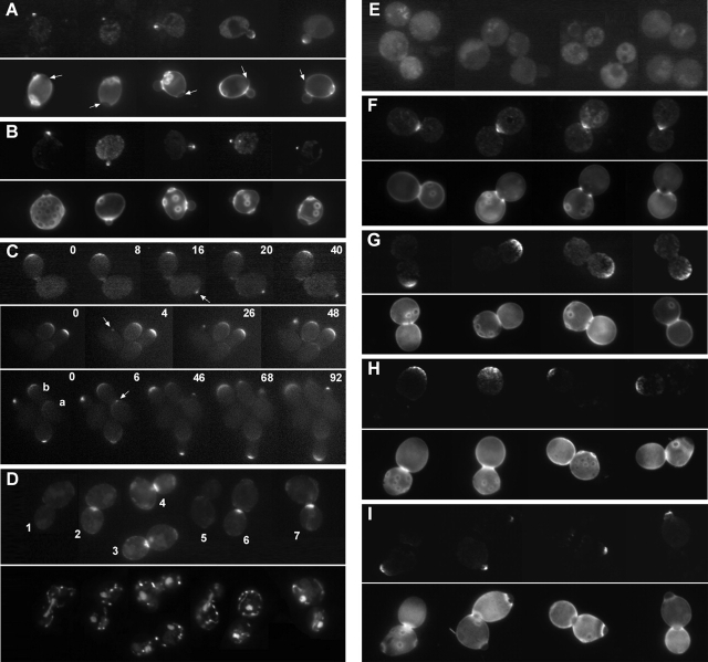
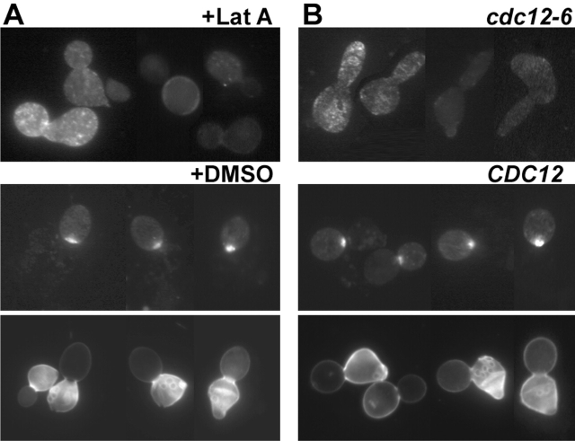
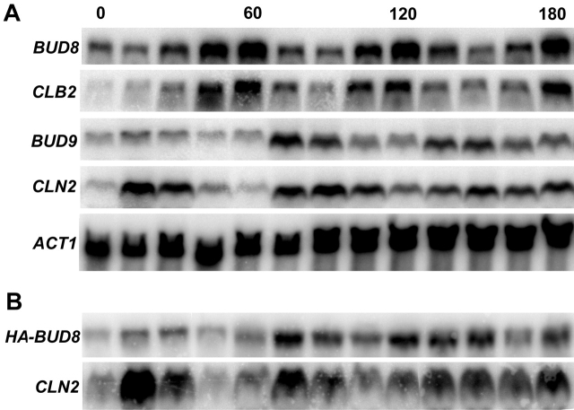
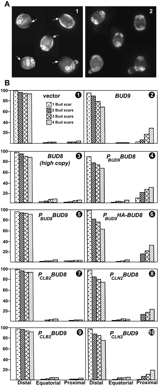
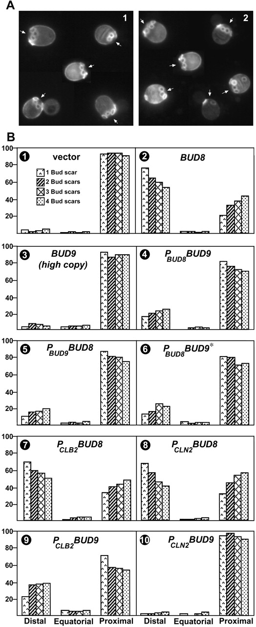
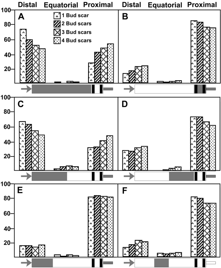
In Saccharomyces cerevisiae, Bud8p and Bud9p are homologous plasma membrane glycoproteins that appear to mark the distal and proximal cell poles, respectively, as potential sites for budding in the bipolar pattern. Here we provide evidence that Bud8p is delivered to the presumptive bud site (and thence to the distal pole of the bud) just before bud emergence, and that Bud9p is delivered to the bud side of the mother-bud neck (and thence to the proximal pole of the daughter cell) after activation of the mitotic exit network, just before cytokinesis. Like the delivery of Bud8p, that of Bud9p is actin dependent; unlike the delivery of Bud8p, that of Bud9p is also septin dependent. Interestingly, although the transcription of BUD8 and BUD9 appears to be cell cycle regulated, the abundance of BUD8 mRNA peaks in G2/M and that of BUD9 mRNA peaks in late G1, suggesting that the translation and/or delivery to the cell surface of each protein is delayed and presumably also cell cycle regulated. The importance of time of transcription in localization is supported by promoter-swap experiments: expression of Bud8p from the BUD9 promoter leads to its localization predominantly to the sites typical for Bud9p, and vice versa. Moreover, expression of Bud8p from the BUD9 promoter fails to rescue the budding-pattern defect of a bud8 mutant but fully rescues that of a bud9 mutant. However, although expression of Bud9p from the BUD8 promoter fails to rescue a bud9 mutant, it also rescues only partially the budding-pattern defect of a bud8 mutant, suggesting that some feature(s) of the Bud8p protein is also important for Bud8p function. Experiments with chimeric proteins suggest that the critical element(s) is somewhere in the extracytoplasmic domain of Bud8p.


Figures

Figure 1.
Figure 1.
Localization of GFP–Bud8p and GFP–Bud9p when expressed from their normal promoters (A and B) or the heterologous promoters (C and D). bud8-Δ1/bud8-Δ1 strain YHH415 (A and C) and bud9-Δ1/bud9-Δ1 strain YHH615 (B and D) were transformed with plasmid YEpGFP*–BUD8F (A), YEpGFP*–BUD9 (B), YEpPBUD8GFP*–BUD9 (C), or YEpPBUD9GFP*–BUD8 (D), grown to exponential phase, stained with Calcofluor as described in Materials and methods, and viewed by fluorescence microscopy for GFP and Calcofluor. In each panel, GFP images are displayed in the top and middle sections, and the bottom section shows the Calcofluor staining of the cells shown in the middle section. The birth scars marking the proximal poles are indicated by arrows. Numbers and asterisks indicate cells discussed in the text.
Figure 2.
Figure 2.
Localization of GFP–Bud8p to bud sites early in the cell cycle and of GFP–Bud9p to the neck late in the cell cycle. bud8-Δ1/bud8-Δ1 strain YHH415 (A and C), rsr1-Δ2/rsr1-Δ2 strain LSY388 (B), and cdc15-2 strain cdc15 (G–I) were transformed with plasmid YEpGFP*–BUD8F; bud9-Δ1/bud9-Δ1 strain HH615 (D) and strain cdc15 (E and F) were transformed with plasmid YEpGFP*–BUD9. (A, B, and D) Cells were grown to exponential phase and stained with Calcofluor (A and B) or DAPI (D) as described in Materials and methods. Individual cells were then imaged for GFP (top section of each panel) and either Calcofluor or DAPI (bottom section of each panel). Arrows indicate birth scars, and numbers indicate cells discussed in the text. (C) Time-lapse observations (Materials and methods) on three groups of cells; times of images (in minutes) are indicated. Note that the quality of focus at particular sites varies from frame to frame. In particular, the presumptive bud site and emerging bud on cell a are well focused at 6, 46, and 92 min, but not at 68 min, whereas the distal pole of cell b is well focused at 6 min and fairly well focused at 46 and 68 min, but out of focus at 92 min. Arrows indicate the nascent bud sites referred to in the text. (E–I) Cells were arrested at 37°C and released as described in Materials and methods. (E and G, top section) GFP images of arrested cells. (G, bottom section) Calcofluor images of cells in the top section. (F, H, and I) GFP (top sections) and Calcofluor (bottom sections) images of individual cells observed at 45 (F and H) or 60 (I) min after release at 23°C.
Figure 3.
Figure 3.
Dependence of GFP–Bud9p localization on actin and the septins. (A) Strain ML130 (MATa bar1Δ) was transformed with YEpGFP*–BUD9, synchronized with α factor, grown for 70 min, and treated for 20 min with Lat A in DMSO or with DMSO alone as described in Materials and methods. (Top and middle panels) GFP images; (bottom panel) Calcofluor images of the cells shown in the middle panel. (B) Strains ML130 and LSY192 (MATa bar1Δ cdc12-6) were transformed with YEpGFP*–BUD9, synchronized with α factor, and then incubated for 90 min at 37°C as described in Materials and methods. (Top and middle panels) GFP images; (bottom panel) Calcofluor images of the cells shown in the middle panel.
Figure 4.
Figure 4.
BUD8 and BUD9 expression through the cell cycle. BUD8 BUD9 strain LSY90 (A) and BUD8 bud9-Δ:: HA-BUD8 strain LSY305 (B) were synchronized with α factor as described in Materials and methods. Total RNAs were extracted from samples taken at 15-min intervals and analyzed by Northern blotting using probes specific for the indicated genes (Materials and methods). Similar results were obtained when RNAs from strain cdc15 were analyzed after synchronization by cdc15 arrest and release (Materials and methods). In B, the use of a probe specific for the HA-epitope sequences allows specific detection of message from the bud9-Δ::HA-BUD8 locus despite the presence of normal BUD8 mRNA.
Figure 5.
Figure 5.
Rescue of a bud9 mutant by either BUD9 or BUD8 if expressed early in the cell cycle. Exponentially growing cells were stained with Calcofluor to evaluate budding patterns. (A) Fluorescence micrographs of bud9-Δ1/bud9-Δ1 strain YHH615 harboring (1) control plasmid YCplac111 or (2) PBUD9BUD8 plasmid YCpPBUD9BUD8. Arrows indicate the birth scars marking the proximal poles. (B) Quantitative evaluation of budding patterns. For each strain, the positions of all bud scars were determined for 100 cells with one bud scar (i.e., 100 total bud scars), 100 cells with two bud scars (i.e., 200 total bud scars), 100 cells with three bud scars (i.e., 300 total bud scars), and 100 cells with four bud scars (i.e., 400 total bud scars). Bud scars were scored as distal (the third of the cell most distal to the birth scar), equatorial (the middle third of the cell), or proximal (the third of the cell surrounding the birth scar). For each strain, the average value from three independent experiments is shown. (1-5 and 7–10) Strain YHH615 harboring plasmid (1) YCplac111, (2) YCpBUD9, (3) YEpBUD8, (4) YCpPBUD9BUD8, (5) YCpPBUD8BUD9, (7) YCpPCLB2BUD8, (8) YCpPCLN2BUD8, (9) YCpPCLB2BUD9, or (10) YCpPCLN2BUD9. (6) bud9-Δ1/bud9-Δ::HA-BUD8 strain LSY492.
Figure 6.
Figure 6.
Rescue of a bud8 mutant by BUD8 if expressed late in the cell cycle and partial rescue by BUD8 if expressed early in the cell cycle or BUD9 if expressed late. Exponentially growing cells were stained with Calcofluor, and budding patterns were evaluated as in Fig. 5. (A) Fluorescence micrographs of bud8-Δ1/bud8-Δ1 strain YHH415 harboring (1) control plasmid YCplac111 or (2) PBUD8BUD9 plasmid YCpPBUD8BUD9. Arrows indicate the birth scars marking the proximal poles. (B) Quantitative evaluation of budding patterns in strain YHH415 harboring plasmid (1) YCplac111, (2) YCpBUD8, (3) YEpBUD9, (4) YCpPBUD8BUD9, (5) YCpPBUD9BUD8, (6) YCpPBUD8BUD9*, (7) YCpPCLB2BUD8, (8) YCpPCLN2BUD8, (9) YCpPCLB2BUD9, or (10) YCpPCLN2BUD9. Plasmids YCpPBUD8BUD9 and YCpPBUD8BUD9* contain downstream sequences from BUD9 and BUD8, respectively.
Figure 7.
Figure 7.
Dependence of Bud8p function on sequences in its extracytoplasmic domain. Exponentially growing cells of bud8-Δ1/bud8-Δ1 strain YHH415 harboring various plasmids were stained with Calcofluor, and budding patterns were evaluated quantitatively as described in Fig. 5. The plasmids contain BUD8/BUD9 chimeras as diagrammed in the figure and described in the Supplemental materials and methods (available at http://www.jcb.org/cgi/content/full/jcb.200107041/DC1). BUD8 regions of the chimeric open reading frames are gray; BUD9 regions are white. Black bars indicate the transmembrane domains. Each chimera contains the BUD8 promoter (gray arrow) and BUD8 (A–E, solid gray line) or BUD9 (F, white line) downstream sequences. Plasmids used were (A) YCpBUD8/9-1, (B) YCpBUD8/9-2, (C) YCpBUD8/9-3, (D) YCpBUD8/9-4, (E) YCpBUD8/9-5, and (F) YCpBUD8/9-6.

Similar articles

Cited by

References

    1. Amberg, D.C., J.E. Zahner, J.W. Mulholland, J.R. Pringle, and D. Botstein. 1997. Aip3p/Bud6p, a yeast actin-interacting protein that is involved in morphogenesis and the selection of bipolar budding sites. Mol. Biol. Cell. 8:729–753. - PMC - PubMed
    1. Ausubel, F.M., R. Brent, R.E. Kingston, D.D. Moore, J.G. Seidman, J.A. Smith, and K. Struhl. 1995. Current Protocols in Molecular Biology. John Wiley & Sons, New York.
    1. Baudin, A., O. Ozier-Kalogeropoulos, A. Denouel, F. Lacroute, and C. Cullin. 1993. A simple and efficient method for direct gene deletion in Saccharomyces cerevisiae. Nucleic Acids Res. 21:3329–3330. - PMC - PubMed
    1. Bender, A., and J.R. Pringle. 1989. Multicopy suppression of the cdc24 budding defect in yeast by CDC42 and three newly identified genes including the ras-related gene RSR1. Proc. Natl. Acad. Sci. USA. 86:9976–9980. - PMC - PubMed
    1. Bi, E., and J.R. Pringle. 1996. ZDS1 and ZDS2, genes whose products may regulate Cdc42p in Saccharomyces cerevisiae. Mol. Cell. Biol. 16:5264–5275. - PMC - PubMed

Publication types

MeSH terms