. 2002 Mar 4;156(5):893-903.
doi: 10.1083/jcb.200107049. Epub 2002 Mar 4.

High RhoA activity maintains the undifferentiated mesenchymal cell phenotype, whereas RhoA down-regulation by laminin-2 induces smooth muscle myogenesis

Affiliations

High RhoA activity maintains the undifferentiated mesenchymal cell phenotype, whereas RhoA down-regulation by laminin-2 induces smooth muscle myogenesis

Safedin Beqaj et al. J Cell Biol. .

Abstract

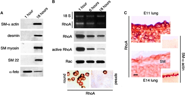
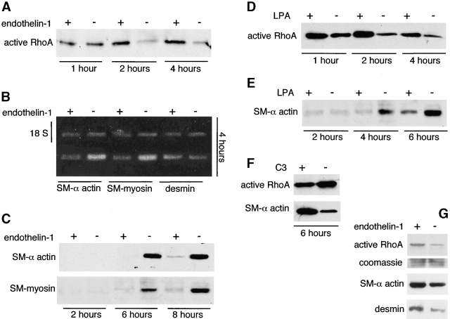
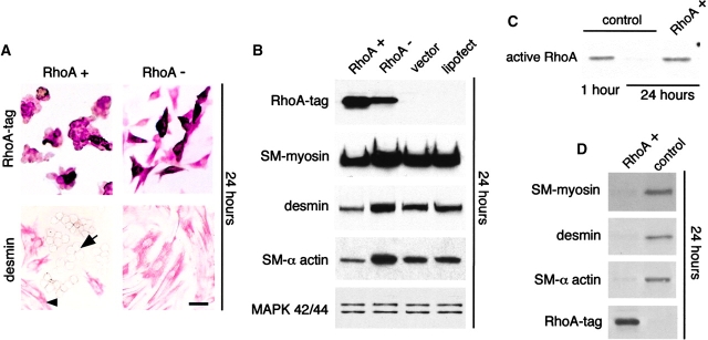
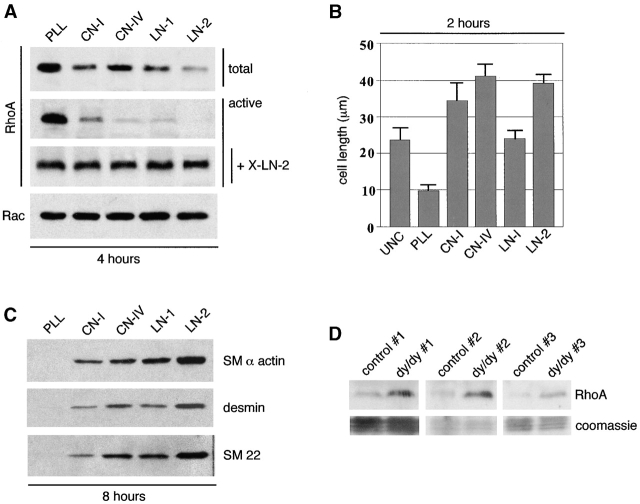
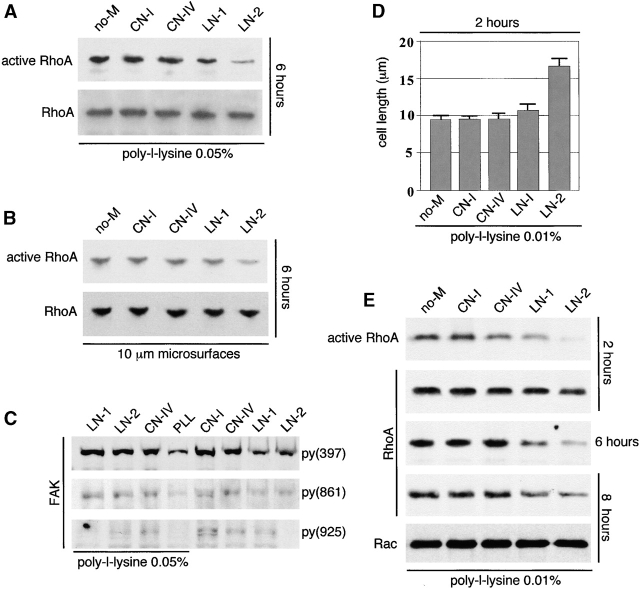
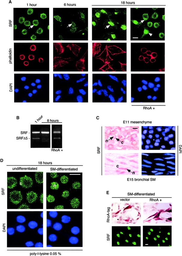
Round embryonic mesenchymal cells have the potential to differentiate into smooth muscle (SM) cells upon spreading/elongation (Yang, Y., K.C. Palmer, N. Relan, C. Diglio, and L. Schuger. 1998. Development. 125:2621-2629; Yang, Y., N.K. Relan, D.A. Przywara, and L. Schuger. 1999. Development. 126:3027-3033; Yang, Y., S. Beqaj, P. Kemp, I. Ariel, and L. Schuger. 2000. J. Clin. Invest. 106:1321-1330). In the developing lung, this process is stimulated by peribronchial accumulation of laminin (LN)-2 (Relan, N.K., Y. Yang, S. Beqaj, J.H. Miner, and L. Schuger. 1999. J. Cell Biol. 147:1341-1350). Here we show that LN-2 stimulates bronchial myogenesis by down-regulating RhoA activity. Immunohistochemistry, immunoblotting, and reverse transcriptase-PCR indicated that RhoA, a small GTPase signaling protein, is abundant in undifferentiated embryonic mesenchymal cells and that its levels decrease along with SM myogenesis. Functional studies using agonists and antagonists of RhoA activation and dominant positive and negative plasmid constructs demonstrated that high RhoA activity was required to maintain the round undifferentiated mesenchymal cell phenotype. This was in part achieved by restricting the localization of the myogenic transcription factor serum response factor (SRF) mostly to the mesenchymal cell cytoplasm. Upon spreading on LN-2 but not on other main components of the extracellular matrix, the activity and level of RhoA decreased rapidly, resulting in translocation of SRF to the nucleus. Both cell elongation and SRF translocation were prevented by overexpression of dominant positive RhoA. Once the cells underwent SM differentiation, up-regulation of RhoA activity induced rather than inhibited SM gene expression. Therefore, our studies suggest a novel mechanism whereby LN-2 and RhoA modulate SM myogenesis.


Figures

Figure 1.
Figure 1.
RhoA expression and activity decrease during SM differentiation in vitro and in vivo. (A and B) Undifferentiated mesenchymal cells isolated from E11 mouse embryonic lungs were cultured for 1 and 18 h. (A) As shown previously (Yang et al., 1999), undifferentiated mesenchymal cells undergo spread-induced SM differentiation. This is in part indicated by the expression of SM-related proteins. In contrast, α-fetoprotein, an embryonic protein, decreases with myogenic cell differentiation. (B) RT-PCR, Western blot analysis, and immunohistochemistry demonstrating rho-a differential expression in embryonic mesenchymal cells undergoing spread-induced SM differentiation in culture. Densitometry analysis showed 8.2-, 7.5-, and 11.5-fold decrease in RhoA mRNA, protein, and activity levels, respectively, after 18 h in culture. No changes were observed in Rac protein levels. Immunohistochemistry, on the bottom left and right, shows high level of cytoplasmic RhoA in round undifferentiated mesenchymal cells and weak, almost negative, RhoA immunostaining in a confluent monolayer of spread SM-differentiating cells. Notice a single cell that remained round and strongly positive for RhoA. (C) RhoA immunolocalization in mouse embryonic lung at days 11 and 14 of gestation. RhoA is diffusely present in the cytoplasm of undifferentiated mesenchymal cells in both gestational stages, although it is more intense at day 11. The elongated bronchial SM cells (SM) show very weak RhoA immunostaining. The small insets represent parallel sections immunostained for SM α-actin to demonstrate absence of SM in E11 lung and its presence in E14 lung. Bars: (C) 10 μm; (insets) 30 μm.
Figure 2.
Figure 2.
Up-regulation of RhoA activity delays SM myogenesis, but once the cells are fully differentiated RhoA stimulates SM gene expression. Undifferentiated mesenchymal cells were isolated from E11 mouse embryonic lungs, allowed to attach, and cultured for up to 8 h in the presence of RhoA agonists endothelin-1 and LPA (A–E) or in the presence of RhoA antagonist C3 (F). Additional cells were allowed to differentiate for 4 d and were then stimulated with endothelin-1 (G). (A) The decrease in RhoA activity normally occurring in mesenchymal cell cultures undergoing spread-induced SM differentiation is prevented in the first hours of culture by 1 μM endothelin-1. These cultures exhibit a significant delay in SM myogenesis as indicated by the lower expression of SM α-actin, desmin, and SM-myosin mRNA (18 S represents the internal control) (B) and protein (C). (D) LPA at a concentration of 10 μM stimulates RhoA activity and as endothelin-1 also inhibits SM myogenesis (E). (F) 10 μM C3 inhibits RhoA activity in the SM-differentiating cultures, and this results in stimulation of SM myogenesis. (G) Once mesenchymal cells differentiate into SM cells after 4 d in culture (Yang et al., 1999), their response to RhoA activation reverses, and then 1 μM endothelin-1 stimulates SM gene expression.
Figure 3.
Figure 3.
Transfection studies with RhoA + and RhoA plasmid constructs, showing that RhoA modulates SM myogenesis by modulating mesenchymal cell shape. Undifferentiated mesenchymal cells were isolated from E11 mouse embryonic lungs and transfected with the two different RhoA plasmid constructs before spreading (1 h after plating). (A) Cells transfected with RhoA+ and immunostained with anti-HA1 tag remain round in shape (top left). Spread nontransfected cells in the same picture cannot be visualized because they are not stained and are in a different focus. Round cells do not immunostain for desmin (bottom left; arrow points to one of these cells). In the same picture, note desmin-positive spread cells (arrowhead points to one of them). Cells transfected with RhoA have spread (top right) and demonstrate strong immunopositivity for desmin (bottom right). (B) Immunoblots of cells transfected with RhoA+ and RhoA. Mesenchymal cells transfected with RhoA spread faster than the nontransfected cells in the same culture, leading to an increase in SM gene expression. For comparison, mitogen-activated protein kinases show no differences in levels. (C and D) RhoA+-transfected mesenchymal cells, which detach faster than spread nontransfected counterparts, were collected from the cultures by short trypsinization and immediately lysed. (C) After 24 h, cells expressing RhoA+ have the same RhoA activity as control undifferentiated mesenchymal cells 1 h after isolation. Here RhoA activity assay was run on 3.5 μg of protein per lane. (D) Cells expressing RhoA+ do not undergo SM myogenesis as indicated by the absence of SM myosin, desmin, and SM α-actin. Note that 3 μg protein/lane was loaded for this experiment rather than the 15 μg protein loaded in other experiments. Thus, the bands seen in the vector control sample are less intense than in other panels. Bar, 20 μm.
Figure 4.
Figure 4.
Mesenchymal cell spreading/elongation on LN-2 causes maximal decrease in the activity and level of RhoA and maximal stimulation of SM myogenesis. (A) Undifferentiated mesenchymal cells were isolated from E11 mouse embryonic lungs and immediately plated on 0.05% poly-l-lysine PLL, CN-1, CN-IV, LN-1, or LN-2 coated onto nontissue culture dishes. RhoA levels and activity decreased rapidly in mesenchymal cells spreading on all of the ECM substrata and particularly on LN-2 but remained high in round cells (plated on PLL). Rac levels were not affected. Since cell elongation per se induces LN-2 synthesis in mesenchymal cells (Relan et al., 1999), 10 μg/ml of anti–LN-2 antibody (X-LN-2) were added to the cultures to block LN-2 activity (Relan et al., 1999). Under these conditions, the cells did not spread and RhoA levels did not decrease. (B) Histogram showing the average length achieved by the cells on different substrata. UNC, uncoated. Note that cells acquired the longest diameters on CN-IV rather than LN-2. Nevertheless, reprobing the membranes shown in A with antibodies to SM myosin, desmin, and SM22 indicates that LN-2 represents the best stimulus for SM myogenesis among the ECM constituents studied (C). (D) RhoA levels in lungs from three dy/dy mice and three matched control animals, showing that dy/dy lungs have significantly higher levels of RhoA. The bottom panels show staining of the blots with Coomassie blue to demonstrate equal protein loading in each set of dy/dy and control lungs.
Figure 5.
Figure 5.
LN-2 down-regulates RhoA activity in the absence of cell elongation. Undifferentiated embryonic mesenchymal cells were isolated from E11 mouse embryonic lungs and plated on poly-l-lysine (A, D, and E) or on 10-μm diameter culture microsurfaces (Yang et al., 1999) (B). CN-I, CN-IV, LN-1, LN-2, or no ECM (no-M) were added to the culture medium once cell attachment was completed (1 h after plating). (A) Cells plated on 0.05% poly-l-lysine, showing that soluble LN-2 decreases RhoA activity. Note that RhoA levels remain unchanged. (B) Mesenchymal cells plated on 10 μm microsurfaces also showing a reduction in RhoA activity without a reduction in RhoA levels upon exposure to LN-2. (C) Immunoblots with three different FAK phosphospecific antibodies, showing that LN-2 elicits low FAK phosphorylation compared with other ECM constituents. (D) Poly-l-lysine was reduced to 0.01% (minimal level required to prevent spontaneous cell spreading). Under these conditions, LN-2 and to a minimal extent LN-1 stimulate mesenchymal cell spreading/elongation. (E) LN-2– and LN-1–induced mesenchymal cell elongation results in down-regulation of both RhoA activity and level.
Figure 6.
Figure 6.
In undifferentiated and immature mesenchymal cells, RhoA activity determines the intracellular localization of SRF. (A) Undifferentiated mesenchymal cells were isolated from E11 mouse embryonic lungs and allowed to undergo spread-induced SM differentiation. Some of the cultures were transfected with RhoA+ before spreading (1 h after plating). Cells were immunostained for SRF 1, 6, and 18 h after plating. In undifferentiated mesenchymal cells, SRF is localized mainly in the cytoplasm (arrow in first panel points to cytoplasm). SRF translocates to the nucleus along with SM differentiation (arrow in second panel points to a cell with nuclear and cytoplasmic SRF, and arrow in third panel points to nuclear SRF). SRF translocation does not occur in undifferentiated mesenchymal cells expressing RhoA+ (right, arrow points to cytoplasm). In same figure, notice presence of SRF in nuclei of spread untransfected cell (arrowhead). Phalloidin stain for F-actin (red) and DAPI stain (blue) are shown to respectively highlight the cytoplasm and nucleus and thereby facilitate interpretation of the figure. (B) RT-PCR shows presence of SRF and SRFΔ5 in undifferentiated mesenchymal cells, presence of SRF but not SRFΔ5 in spread cells 8 h after plating, and presence of both in RhoA+-transfected cells. (C) Immunohistochemical studies performed on E11 and E15 embryonic lung sections, revealing cytoplasmic localization of SRF in undifferentiated mesenchymal cells in vivo (top, arrow points to cytoplasm) and its nuclear localization in SM-differentiated cells in vivo (bottom, arrow points to nucleus). n, nucleus; c, cytoplasm. (D) Undifferentiated mesenchymal cells were cultured for either 1 or 24 h. The cells were then trypsinized, replated on poly-l-lysine, and cultured for another 18 h. At the end of the second culture period, the cells were immunostained for SRF. Although cells replated on poly-l-lysine after 1 h in regular culture still have most SRF immunoreactivity in the cytoplasm (left; c, cytoplasm), cells that were allowed to spread for 24 h before replating on poly-l-lysine have all SRF immunoreactivity in the nucleus (right; n, nucleus). (E) Spread SM-differentiating mesenchymal cells transfected with RhoA+ or vector alone and immunostained with anti-HA1 tag by immunoperoxidase (top) or with anti-SRF by immunofluorescence (bottom). Immunostaining for SRF shows no translocation of SRF to the cytoplasm. Notice that although we were unable to develop a double immunostaining for SRF and HA1-tagged RhoA, since ∼25–30% of the cells were transfected we should have been able to detect scattered cells with changes in SRF localization if such changes had taken place. Bars, 10 μm.

Similar articles

Cited by

References

    1. Adams, J.C., and M.A. Schwartz. 2000. Stimulation of fascin spikes by thrombospondin-1 is mediated by the GTPases Rac and Cdc42. J. Cell Biol. 150:807–822. - PMC - PubMed
    1. Belaguli, N.S., L.A. Schildmeyer, and R.J. Schwartz. 1997. Organization and myogenic restricted expression of the murine serum response factor gene. A role for autoregulation. J. Biol. Chem. 272:18222–18231. - PubMed
    1. Belaguli, N.S., W. Zhou, T.H. Trinh, M.W. Majesky, and R.J. Schwartz. 1999. Dominant negative murine serum response factor: alternative splicing within the activation domain inhibits transactivation of serum response factor binding targets. Mol. Cell. Biol. 19:4582–4591. - PMC - PubMed
    1. Berkowitz, L.A., K.T. Riabowol, and M.Z. Gilman. 1989. Multiple sequence elements of a single functional class are required for cyclic AMP responsiveness of the mouse c-fos promoter. Mol. Cell. Biol. 9:4272–4281. - PMC - PubMed
    1. Bishop, A.L., and A. Hall. 2000. Rho GTPases and their effector proteins. Biochem. J. 348:241–255. - PMC - PubMed

Publication types

MeSH terms