

Review
doi: 10.1172/JCI15198.
Degeneracy, as opposed to specificity, in immunotherapy
Affiliations
- PMID: 11877465
- PMCID: PMC150898
- DOI: 10.1172/JCI15198
Item in Clipboard
Review
Degeneracy, as opposed to specificity, in immunotherapy
J Clin Invest.
2002 Mar.
No abstract available
Figures
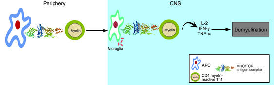
Autoimmune mechanism of MS. Naive, myelin reactive T cells are activated by microbes that: 1) induce the innate immune system to provide costimulatory signals necessary for clonal expansion of naive CD4+ T cells and, 2) contain epitopes that are cross-reactive with self antigens. Activated CD4+ T cells cross the blood-brain barrier and recognize self antigen presented by microglia, local antigen-presenting cells (APC) that sample self antigens. The reactivation of autoreactive CD4+ cells leads to a cascade of events including recruitment of macrophages, antibody secretion, and eventual destruction of myelin and axons.
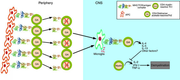
The CD4+ T cell model of action by glatiramer acetate (GA). GA induces strong MHC class II restricted proliferative responses by T cells. Daily injections of GA induces a moderate loss of responsiveness to the antigen accompanied by a shift to a more Th2 type of CD4+ T cell. The surviving GA-reactive T cells show a greater degree of degeneracy, as measured by cross-reactive responses to combinatorial peptide libraries. Highly cross-reactive Th2 cells with degenerate T cell receptors migrate to the site CNS, recognize self antigens as weak agonists or “altered peptide ligands”. In response, they begin to secrete Th2/Th3 cytokines, and suppress inflammation by the mechanism of bystander suppression.
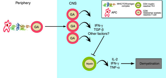
The CD8+ T cell model of action by GA. MHC class I restricted CD8+ T cell responses to GA, which are significantly lower at baseline in MS patients than in healthy controls, increase significantly following treatment, as seen in the greater number of IFN-γ–positive, GA responsive CD8+ T cells. GA treatment is proposed to restore CD8+ suppressor function, which is dependent upon IFN-γ secretion. The mechanism for CD8+ suppression of the immune response is unknown. Shown in the figure is a CD8+ T cell migrating to the CNS, regulating a CD4+ autoreactive T cell.
Comment on
-
Glatiramer acetate (Copaxone) therapy induces CD8(+) T cell responses in patients with multiple sclerosis.J Clin Invest. 2002 Mar;109(5):641-9. doi: 10.1172/JCI14380. J Clin Invest. 2002. PMID: 11877472 Free PMC article.
Similar articles
-
CD8+ T cells maintain tolerance to myelin basic protein by 'epitope theft'.Nat Immunol. 2004 Jun;5(6):606-14. doi: 10.1038/ni1073. Epub 2004 May 16. Nat Immunol. 2004. PMID: 15146180
-
Blood CD8+ T cell responses against myelin determinants in multiple sclerosis and healthy individuals.Eur J Immunol. 2008 Jul;38(7):1889-99. doi: 10.1002/eji.200838023. Eur J Immunol. 2008. PMID: 18506883
-
Activation of myelin reactive T cells in multiple sclerosis: a possible role for T cell degeneracy?Eur J Immunol. 2008 May;38(5):1190-3. doi: 10.1002/eji.200838371. Eur J Immunol. 2008. PMID: 18425726
-
Specificity and degeneracy: T cell recognition in CNS autoimmunity.Mol Immunol. 2004 Feb;40(14-15):1057-61. doi: 10.1016/j.molimm.2003.11.012. Mol Immunol. 2004. PMID: 15036910 Review.
-
Specificity and degeneracy of T cells.Mol Immunol. 2004 Feb;40(14-15):1047-55. doi: 10.1016/j.molimm.2003.11.022. Mol Immunol. 2004. PMID: 15036909 Review.
Cited by
-
Hit-Hit and hit-Run: viruses in the playing field of multiple sclerosis.Curr Neurol Neurosci Rep. 2003 May;3(3):265-71. doi: 10.1007/s11910-003-0087-9. Curr Neurol Neurosci Rep. 2003. PMID: 12760396 Review.
-
The good and the bad of T cell cross-reactivity: challenges and opportunities for novel therapeutics in autoimmunity and cancer.Front Immunol. 2023 Jun 19;14:1212546. doi: 10.3389/fimmu.2023.1212546. eCollection 2023. Front Immunol. 2023. PMID: 37409132 Free PMC article. Review.
-
T-cell-based vaccination for morphological and functional neuroprotection in a rat model of chronically elevated intraocular pressure.J Mol Med (Berl). 2005 Nov;83(11):904-16. doi: 10.1007/s00109-005-0689-6. Epub 2005 Aug 12. J Mol Med (Berl). 2005. PMID: 16096740
-
Interferon-beta therapy reduces CD4+ and CD8+ T-cell reactivity in multiple sclerosis.Immunology. 2007 May;121(1):29-39. doi: 10.1111/j.1365-2567.2006.02518.x. Epub 2006 Dec 18. Immunology. 2007. PMID: 17239199 Free PMC article.
-
Glatiramer acetate treatment effects on gene expression in monocytes of multiple sclerosis patients.J Neuroinflammation. 2013 Oct 17;10:126. doi: 10.1186/1742-2094-10-126. J Neuroinflammation. 2013. PMID: 24134771 Free PMC article.
References
-
- Ota K, et al. T-cell recognition of an immunodominant myelin basic protein epitope in multiple sclerosis. Nature. 1990;346:183–187. - PubMed
