

Overexpression of the serpin megsin induces progressive mesangial cell proliferation and expansion
- PMID: 11877466
- PMCID: PMC150894
- DOI: 10.1172/JCI14336
Overexpression of the serpin megsin induces progressive mesangial cell proliferation and expansion
Abstract
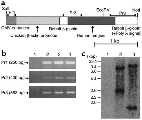
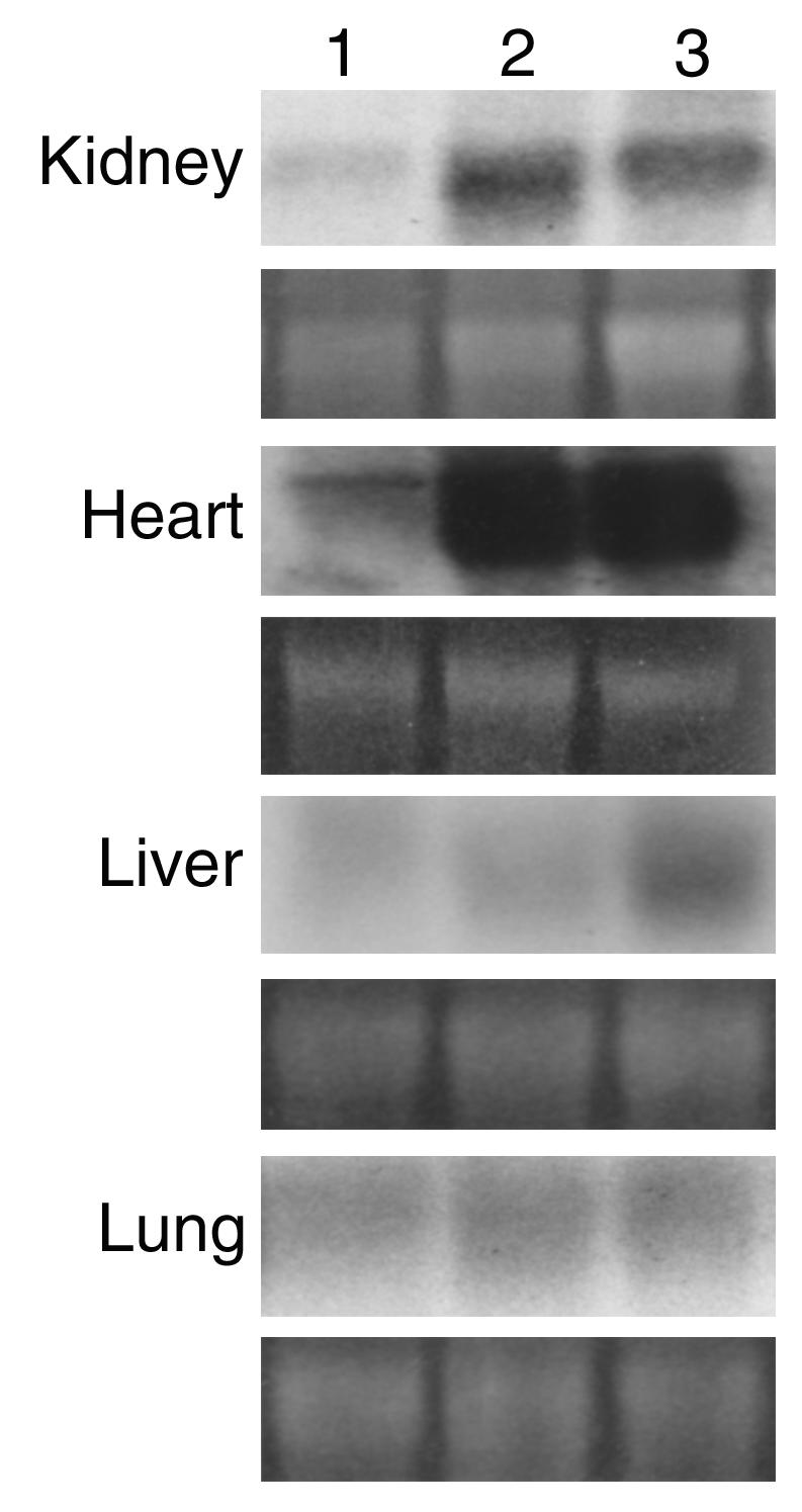
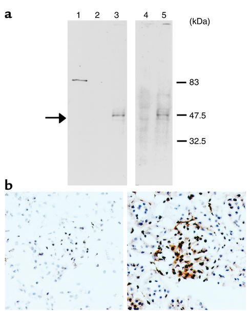
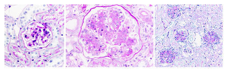
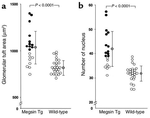
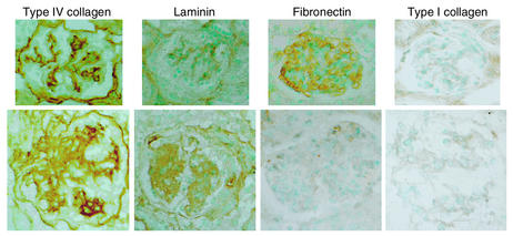
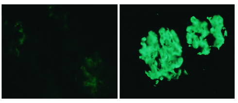
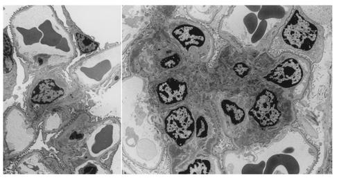
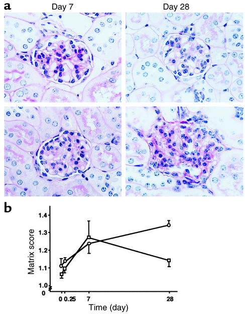
Mesangial cells maintain normal glomerular function by mediating ECM remodeling and immune complex disposal. We have recently identified megsin, a novel member of the serine protease inhibitor (serpin) superfamily predominantly expressed in the mesangium. While our previous studies suggested a role for megsin in the pathogenesis of human glomerular diseases, its exact biological significance remained unknown. Here we produced two lines of megsin transgenic mice. Overexpression of megsin led to progressive mesangial matrix expansion and an increase in the number of mesangial cells. These glomerular lesions were accompanied by an augmented immune complex deposition, together with Ig's and complement. Binding and functional assays in vitro identified plasmin as one biological substrate of megsin and confirmed its activity as a proteinase inhibitor. Transgenic animals exhibiting nephritis as a result of treatment with anti--glomerular basement membrane antiserum showed significantly more persistent expansion of the mesangial ECM than was seen in parental mice. Megsin therefore exerts a biologically relevant influence on mesangial function, and on the mesangial microenvironment, such that simple overexpression of this endogenous serpin engenders elementary mesangial lesions.
Figures
Similar articles
-
Accelerated glomerular injury in hemi-nephrectomized transgenic mice of mesangial cell-predominant serpin, megsin.Nephron Exp Nephrol. 2004;96(4):e127-33. doi: 10.1159/000077379. Nephron Exp Nephrol. 2004. PMID: 15122062
-
Mesangial cell-predominant gene, megsin.Nephrol Dial Transplant. 2002;17 Suppl 9:32-3. doi: 10.1093/ndt/17.suppl_9.32. Nephrol Dial Transplant. 2002. PMID: 12386281
-
Mesangial cell-predominant functional gene, megsin.Clin Exp Nephrol. 2003 Jun;7(2):87-92. doi: 10.1007/s10157-003-0228-0. Clin Exp Nephrol. 2003. PMID: 14586725 Review.
-
Expression of megsin mRNA, a novel mesangium-predominant gene, in the renal tissues of various glomerular diseases.J Am Soc Nephrol. 1999 Dec;10(12):2606-13. doi: 10.1681/ASN.V10122606. J Am Soc Nephrol. 1999. PMID: 10589701
-
Proteomics and mesangial cell: serpin, megsin and plasmin.Contrib Nephrol. 2004;141:212-20. doi: 10.1159/000074599. Contrib Nephrol. 2004. PMID: 14650234 Review. No abstract available.
Cited by
-
Bomapin is a redox-sensitive nuclear serpin that affects responsiveness of myeloid progenitor cells to growth environment.BMC Cell Biol. 2010 Apr 30;11:30. doi: 10.1186/1471-2121-11-30. BMC Cell Biol. 2010. PMID: 20433722 Free PMC article.
-
SerpinB7 deficiency contributes to development of psoriasis via calcium-mediated keratinocyte differentiation dysfunction.Cell Death Dis. 2022 Jul 21;13(7):635. doi: 10.1038/s41419-022-05045-8. Cell Death Dis. 2022. PMID: 35864103 Free PMC article.
-
Megsin gene: its genomic analysis, pathobiological functions, and therapeutic perspectives.Curr Genomics. 2007 May;8(3):203-8. doi: 10.2174/138920207780833856. Curr Genomics. 2007. PMID: 18645605 Free PMC article.
-
Systematic identification and characterization of novel human skin-associated genes encoding membrane and secreted proteins.PLoS One. 2013 Jun 20;8(6):e63949. doi: 10.1371/journal.pone.0063949. Print 2013. PLoS One. 2013. PMID: 23840300 Free PMC article.
-
Large-scale identification of human genes implicated in epidermal barrier function.Genome Biol. 2007;8(6):R107. doi: 10.1186/gb-2007-8-6-r107. Genome Biol. 2007. PMID: 17562024 Free PMC article.
References
-
- Suzuki D, et al. Expression of megsin mRNA, a novel mesangium-predominant gene, in the renal tissues of various glomerular diseases. J Am Soc Nephrol. 1999;10:2606–2613. - PubMed
-
- Inagi R, et al. Specific tissue distribution of megsin, a novel serpin, in the glomerulus and its up-regulation in IgA nephropathy. Biochem Biophys Res Commun. 2001;286:1098–1106. - PubMed
-
- Nangaku M, et al. Cloning of rodent megsin revealed its up-regulation in mesangioproliferative nephritis. Kidney Int. 2001;60:641–652. - PubMed
-
- Kawarabayashi T, et al. Accumulation of β-amyloid fibrils in pancreas of transgenic mice. Neurobiol Aging. 1996;17:215–222. - PubMed
Publication types
MeSH terms
Substances
LinkOut - more resources
Full Text Sources
Other Literature Sources
Molecular Biology Databases
