

CCR3 is essential for skin eosinophilia and airway hyperresponsiveness in a murine model of allergic skin inflammation
- PMID: 11877470
- PMCID: PMC150891
- DOI: 10.1172/JCI14097
CCR3 is essential for skin eosinophilia and airway hyperresponsiveness in a murine model of allergic skin inflammation
Abstract
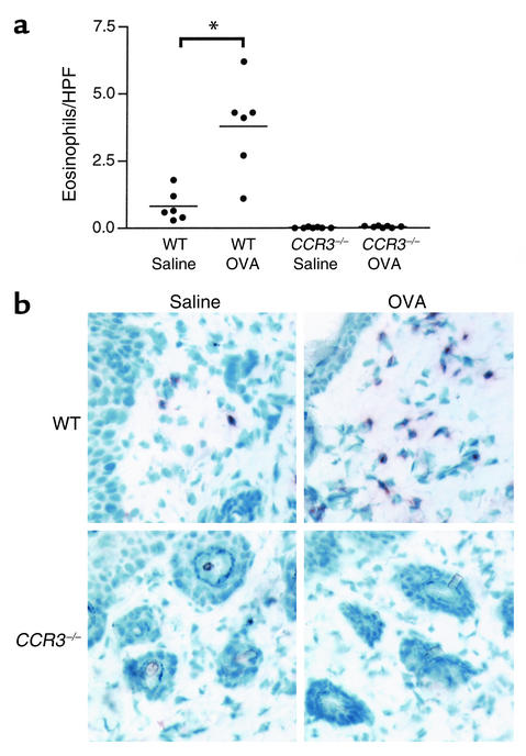
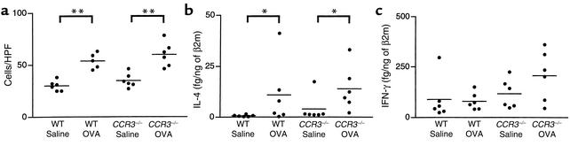
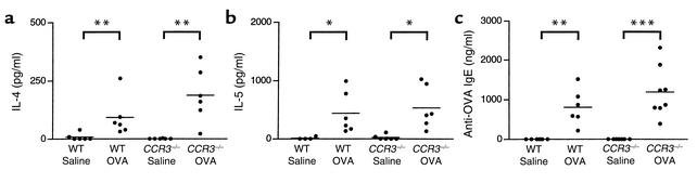
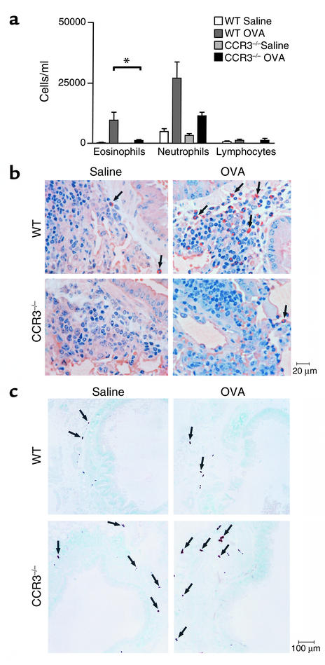
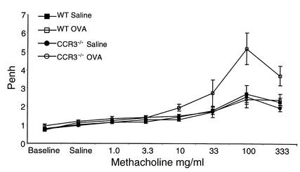
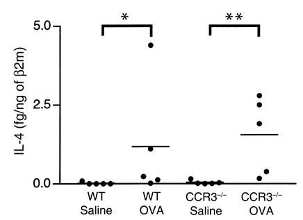
The CC chemokine receptor 3 (CCR3) is expressed by eosinophils, mast cells, and Th2 cells. We used CCR3(-/-) mice to assess the role of CCR3 in a murine model of allergic skin inflammation induced by repeated epicutaneous sensitization with ovalbumin (OVA), and characterized by eosinophil skin infiltration, local expression of Th2 cytokines, and airway hyperresponsiveness (AHR) to inhaled antigen. Eosinophils and the eosinophil product major basic protein were absent from the skin of sham and OVA-sensitized CCR3(-/-) mice. Mast cell numbers and expression of IL-4 mRNA were normal in skin of CCR3(-/-) mice, suggesting that CCR3 is not important for infiltration of the skin by mast cells and Th2 cells. CCR3(-/-) mice produced normal levels of OVA-specific IgE, and their splenocytes secreted normal amounts of IL-4 and IL-5 following in vitro stimulation with OVA, indicating effective generation of systemic Th2 helper responses. Recruitment of eosinophils to lung parenchyma and bronchoalveolar lavage (BAL) fluid was severely impaired in CCR3(-/-) mice, which failed to develop AHR to methacholine following antigen inhalation. These results suggest that CCR3 plays an essential role in eosinophil recruitment to the skin and the lung and in the development of AHR.
Figures
Similar articles
-
Exaggerated IL-17 response to epicutaneous sensitization mediates airway inflammation in the absence of IL-4 and IL-13.J Allergy Clin Immunol. 2009 Oct;124(4):761-70.e1. doi: 10.1016/j.jaci.2009.07.040. J Allergy Clin Immunol. 2009. PMID: 19815118 Free PMC article.
-
IL-22 promotes allergic airway inflammation in epicutaneously sensitized mice.J Allergy Clin Immunol. 2019 Feb;143(2):619-630.e7. doi: 10.1016/j.jaci.2018.05.032. Epub 2018 Jun 18. J Allergy Clin Immunol. 2019. PMID: 29920352 Free PMC article.
-
The murine CCR3 receptor regulates both the role of eosinophils and mast cells in allergen-induced airway inflammation and hyperresponsiveness.Proc Natl Acad Sci U S A. 2002 Feb 5;99(3):1479-84. doi: 10.1073/pnas.261462598. Proc Natl Acad Sci U S A. 2002. PMID: 11830666 Free PMC article.
-
The role of human mast cell-derived cytokines in eosinophil biology.J Interferon Cytokine Res. 2004 May;24(5):271-81. doi: 10.1089/107999004323065057. J Interferon Cytokine Res. 2004. PMID: 15153310 Review.
-
Role of IgE in the development of allergic airway inflammation and airway hyperresponsiveness--a murine model.Allergy. 1999 Apr;54(4):297-305. doi: 10.1034/j.1398-9995.1999.00085.x. Allergy. 1999. PMID: 10371087 Review.
Cited by
-
Experimental advances in understanding allergic airway inflammation.Front Biosci (Schol Ed). 2013 Jan 1;5(1):167-80. doi: 10.2741/s364. Front Biosci (Schol Ed). 2013. PMID: 23277043 Free PMC article. Review.
-
Effects of a dual CCR3 and H1-antagonist on symptoms and eosinophilic inflammation in allergic rhinitis.Respir Res. 2010 Feb 9;11(1):17. doi: 10.1186/1465-9921-11-17. Respir Res. 2010. PMID: 20144207 Free PMC article. Clinical Trial.
-
The anaphylatoxin C3a downregulates the Th2 response to epicutaneously introduced antigen.J Clin Invest. 2004 Aug;114(3):399-407. doi: 10.1172/JCI19082. J Clin Invest. 2004. PMID: 15286806 Free PMC article.
-
CCL24 regulates biliary inflammation and fibrosis in primary sclerosing cholangitis.JCI Insight. 2023 Jun 22;8(12):e162270. doi: 10.1172/jci.insight.162270. JCI Insight. 2023. PMID: 37345655 Free PMC article.
-
Anti-IL5 decreases the number of eosinophils but not the severity of dermatitis in Sharpin-deficient mice.Exp Dermatol. 2010 Mar;19(3):252-8. doi: 10.1111/j.1600-0625.2009.00944.x. Epub 2009 Jul 23. Exp Dermatol. 2010. PMID: 19650867 Free PMC article.
References
-
- Leung DYM. Atopic dermatitis: the skin as a window into the pathogenesis of chronic allergic disease. J Allergy Clin Immunol. 1995;96:312–319. - PubMed
-
- Leung DYM. Immunopathology of atopic dermatitis. Springer Semin Immunopathol. 1992;13:427–440. - PubMed
-
- Thepen T, et al. Biphasic response against aeroallergen in atopic dermatitis showing a switch from an initial Th2 response to a Th1 response in situ: an immunocytochemical study. J Allergy Clin Immunol. 1996;97:828–837. - PubMed
-
- Leung DY. Atopic dermatitis: new insights and opportunities for therapeutic intervention. J Allergy Clin Immunol. 2000;105:860–876. - PubMed
-
- Gleich GJ. Mechanisms of eosinophil-associated inflammation. J Allergy Clin Immunol. 2000;105:651–663. - PubMed
Publication types
MeSH terms
Substances
Grants and funding
LinkOut - more resources
Full Text Sources
Other Literature Sources
Medical
Molecular Biology Databases
