. 2002 Mar;109(5):661-70.
doi: 10.1172/JCI13572.

Elastase-mediated phosphatidylserine receptor cleavage impairs apoptotic cell clearance in cystic fibrosis and bronchiectasis

Affiliations

Elastase-mediated phosphatidylserine receptor cleavage impairs apoptotic cell clearance in cystic fibrosis and bronchiectasis

R William Vandivier et al. J Clin Invest. 2002 Mar.

Abstract

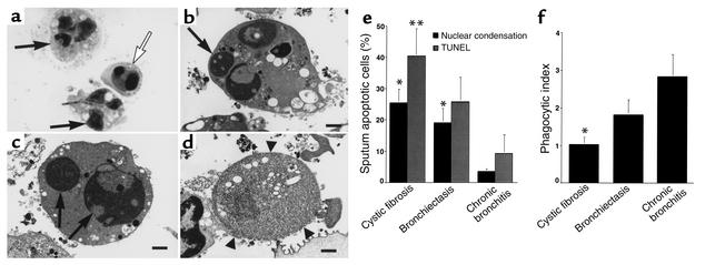
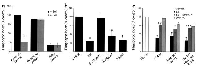
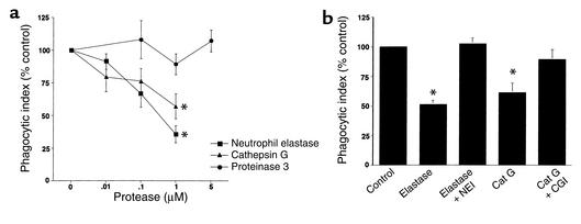
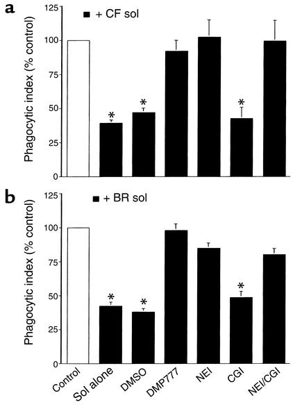
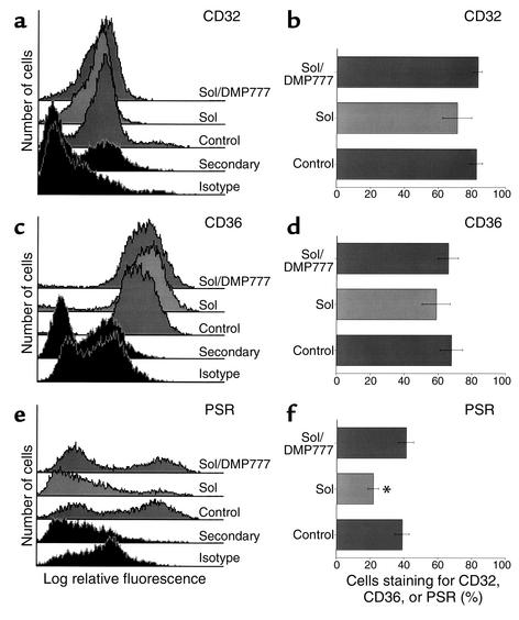
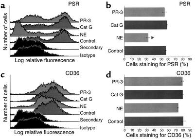
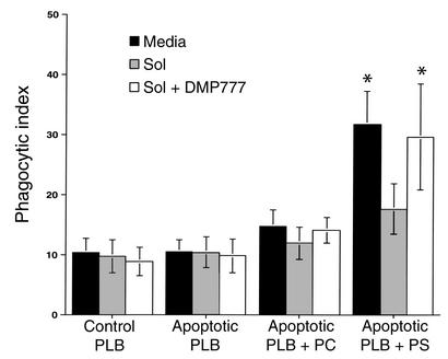
Cystic fibrosis is characterized by an early and sustained influx of inflammatory cells into the airways and by release of proteases. Resolution of inflammation is normally associated with the orderly removal of dying apoptotic inflammatory cells through cell recognition receptors, such as the phosphatidylserine receptor, CD36, and alpha v integrins. Accordingly, removal of apoptotic inflammatory cells may be impaired in persistent inflammatory responses such as that seen in cystic fibrosis airways. Examination of sputa from cystic fibrosis and non-cystic fibrosis bronchiectasis patients demonstrated an abundance of apoptotic cells, in excess of that seen in patients with chronic bronchitis. In vitro, cystic fibrosis and bronchiectasis airway fluid directly inhibited apoptotic cell removal by alveolar macrophages in a neutrophil elastase-dependent manner, suggesting that elastase may impair apoptotic cell clearance in vivo. Flow cytometry demonstrated that neutrophil elastase cleaved the phosphatidylserine receptor, but not CD36 or CD32 (Fc gamma RII). Cleavage of the phosphatidylserine receptor by neutrophil elastase specifically disrupted phagocytosis of apoptotic cells, implying a potential mechanism for delayed apoptotic cell clearance in vivo. Therefore, defective airway clearance of apoptotic cells in cystic fibrosis and bronchiectasis may be due to elastase-mediated cleavage of phosphatidylserine receptor on phagocytes and may contribute to ongoing airway inflammation.


Figures

Figure 1
Figure 1
Clearance of apoptotic cells is defective in CF airways. (a) Wright’s Giemsa stain of CF sputum (×100) showing apoptotic cells with condensed nuclei (open arrow) and normal PMNs (arrows). (bd) Transmission electron micrograph of sputum from a CF patient (×6,300; bars: 1 μm) demonstrating early apoptosis (b), late apoptosis (c), and postapoptotic necrosis (d). Arrows (b and c) indicate condensed apoptotic nuclei. Arrowheads (d) show loss of membrane integrity during necrosis. (e) Sputa from CF or bronchiectasis patients contain more apoptotic cells compared with chronic bronchitis. The percentage of apoptotic cells (by nuclear condensation and TUNEL staining) ± SEM is shown for six patients per group. *Apoptosis by nuclear condensation is significantly different from chronic bronchitis (P < 0.05). **Apoptosis by TUNEL staining is significantly different from chronic bronchitis (P < 0.05). (f) Airway macrophages from CF patients ingest fewer apoptotic cells. The mean phagocytic index of sputum macrophages ± SEM is shown for six patients per group. *Phagocytic index is significantly different from chronic bronchitis (P < 0.05).
Figure 2
Figure 2
CF sol inhibits HMDM ingestion of apoptotic but not IgG-opsonized Jurkat cells in an elastase-dependent manner. (a) HMDMs were cocultured with apoptotic, opsonized, or viable Jurkat cells in the presence or absence of 10% CF sol. The mean phagocytic index as percentage of control ± SEM is shown for three replicates per group. Control mean phagocytic index: 28.6 ± 4.4. *Significantly different from apoptotic Jurkat cells pretreated with media (P < 0.05). (b) HMDMs were cocultured with apoptotic Jurkat cells in the presence of media (Control), 10% CF sol, 10% CF sol plus DMP777 (100 μM), 10% CF sol plus SJ527 (100 μM), or 10% CF sol plus methyl cellulose (MC). The mean phagocytic index as percentage of control ± SEM is shown for three to five replicates per group. Control mean phagocytic index: 52.8 ± 10.2. *Significantly different from control (P < 0.05). (c) HMDMs, apoptotic Jurkat cells, or both were pretreated for 30 minutes with media (Control), 10% CF sol, 10% CF sol plus DMP777 (20 μM), or DMP777 (20 μM) alone prior to coculture. Following pretreatment, coculture was done in the presence of DMP777 (20 μM) to prevent carryover of elastase activity. The mean phagocytic index as percentage of control ± SEM is shown for three replicates per group. Control mean phagocytic index: 51.4 ± 6.0. *Significantly different from control (P < 0.05). **Significantly different from 10% CF sol-pretreated HMDMs (P < 0.05). ***Significantly different from 10% CF sol-pretreated HMDMs and apoptotic Jurkat cells (P < 0.05).
Figure 3
Figure 3
Elastase, or cathepsin G, inhibits ingestion of apoptotic cells. (a) HMDMs were cocultured with apoptotic Jurkat cells in the presence of media or increasing concentrations of elastase, cathepsin G, or proteinase 3. The mean phagocytic index as percentage of control ± SEM is shown for three to six replicates per group. Control mean phagocytic index: 44.1 ± 8.5. *Significantly different from media (P < 0.05). (b) Human AMs were cocultured with apoptotic human PMNs following pretreatment with media (Control), 1 μM PMN elastase (NE) with or without a specific elastase inhibitor (NEI, 100 μM), or 1 μM cathepsin G (Cat G) with or without a specific CGI (10 μM). Coculture was done in the presence of DMP777 (20 μM) to prevent carryover of elastase activity. The mean phagocytic index as percentage of control ± SEM is shown for three or four replicates per group. Control mean phagocytic index: 8.4 ± 1.9. *Significantly different from control (P < 0.05).
Figure 4
Figure 4
CF and non-CF bronchiectasis sol-derived elastase disrupts human AM ingestion of apoptotic human PMNs. Human AMs were cocultured with apoptotic human PMNs following pretreatment with media (Control), 10% CF (a), or 10% non-CF bronchiectasis (BR) (b) sol with or without DMSO, DMP777 (20 μM), NEI (100 μM), CGI (10 μM), or NEI and CGI. Coculture was done in the presence of DMP777 (20 μM) to prevent carryover of elastase activity. The mean phagocytic index as percentage of control ± SEM is shown for three to four replicates per group. Control mean phagocytic index: (a) 7.9 ± 1.7; (b) 7.7 ± 1.4. *Groups indicated were significantly different from control (P < 0.05).
Figure 5
Figure 5
CF sol cleaves PS receptor (PSR) in an elastase-dependent manner. HMDMs were incubated with media (Control), 10% CF sol, or 10% CF sol plus DMP777 (20 μM) for 2 hours. Flow cytometry was done to assess surface staining for CD32 (a and b), CD36 (c and d), and PSR (e and f). Histograms for each receptor are shown in the left panels, and the percent of HMDMs that stain for each receptor ± SEM are shown in the right panels (four to six replicates per group). *Significantly different from control (P < 0.05).
Figure 6
Figure 6
Elastase, but not cathepsin G or proteinase 3, cleaves cell surface PSR. HMDMs were incubated with media (Control), elastase (1 μM), cathepsin G (1 μM), or proteinase 3 (1 μM) for 2 hours. Flow cytometry was done to assess surface staining for PS receptor (a and b) and CD36 (c and d). Histograms for each receptor are shown in the left panels, and the percent of HMDMs that stain for each receptor ± SEM are shown in the right panels (three replicates per group). *Significantly different from control (P < 0.05).
Figure 7
Figure 7
CF sol interferes with PS-mediated apoptotic cell ingestion in an elastase-dependent manner. HMDMs were pretreated with media, 10% CF sol, or 10% CF sol plus DMP777 (20 μM) for 30 minutes prior to coculture with viable or apoptotic PLB985 cells. PS or PC was inserted into the outer membrane of some apoptotic PLB985 cells by phospholipid transfer. HMDMs were then cocultured for 1 hour with viable PLB985 cells (Control), apoptotic PLB985 cells, apoptotic PLB985 cells plus PC, or apoptotic PLB985 cells plus PS. Coculture was done in the presence of DMP777 (20 μM) to prevent carryover of elastase activity. The mean phagocytic index ± SEM is shown for five replicates per group. *Significantly different from control (P < 0.05).

References

    1. Wyllie AH, Kerr JF, Currie AR. Cell death: the significance of apoptosis. Int Rev Cytol. 1980;68:251–306. - PubMed
    1. J.S. Savill, et al. Macrophage phagocytosis of aging neutrophils in inflammation. Programmed cell death in the neutrophil leads to its recognition by macrophages. J Clin Invest. 1989;83:865–875. - PMC - PubMed
    1. Lethem MI, James SL, Marriott C, Burke JF. The origin of DNA associated with mucus glycoproteins in cystic fibrosis sputum. Eur Respir J. 1990;3:19–23. - PubMed
    1. Fadok VA, et al. Macrophages that have ingested apoptotic cells in vitro inhibit proinflammatory cytokine production through autocrine/paracrine mechanisms involving TGF-beta, PGE2, and PAF. J Clin Invest. 1998;101:890–898. - PMC - PubMed
    1. McDonald PP, Fadok VA, Bratton D, Henson PM. Transcriptional and translational regulation of inflammatory mediator production by endogenous TGF-beta in macrophages that have ingested apoptotic cells. J Immunol. 1999;163:6164–6172. - PubMed

Publication types