. 2002 Mar 4;195(5):547-58.
doi: 10.1084/jem.20011465.

Reciprocal roles for CCAAT/enhancer binding protein (C/EBP) and PU.1 transcription factors in Langerhans cell commitment

Affiliations

Reciprocal roles for CCAAT/enhancer binding protein (C/EBP) and PU.1 transcription factors in Langerhans cell commitment

Atsushi Iwama et al. J Exp Med. .

Abstract

Myeloid progenitor cells give rise to a variety of progenies including dendritic cells. However, the mechanism controlling the diversification of myeloid progenitors into each progeny is largely unknown. PU.1 and CCAAT/enhancing binding protein (C/EBP) family transcription factors have been characterized as key regulators for the development and function of the myeloid system. However, the roles of C/EBP transcription factors have not been fully identified because of functional redundancy among family members. Using high titer--retroviral infection, we demonstrate that a dominant-negative C/EBP completely blocked the granulocyte--macrophage commitment of human myeloid progenitors. Alternatively, Langerhans cell (LC) commitment was markedly facilitated in the absence of tumor necrosis factor (TNF)alpha, a strong inducer of LC development, whereas expression of wild-type C/EBP in myeloid progenitors promoted granulocytic differentiation, and completely inhibited TNFalpha-dependent LC development. On the other hand, expression of wild-type PU.1 in myeloid progenitors triggered LC development in the absence of TNFalpha, and its instructive effect was canceled by coexpressed C/EBP. Our findings establish reciprocal roles for C/EBP and PU.1 in LC development, and provide new insight into the molecular mechanism of LC development, which has not yet been well characterized.


Figures

Figure 1.
Figure 1.
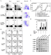
Altered differentiation of CD34+ progenitors expressing a dominant-negative C/EBP. (A) The schematic representation of the retroviral vector, GCsam–A-C/EBP–IRES–EGFP, encoding A-C/EBP, a dominant-negative C/EBP, linked by an IRES to a cDNA encoding EGFP. The 3′ LTR of the vector is replaced with MSCV. ψ+, packaging signal; SD, splice donor; SA, splice acceptor. (B) The effect of A-C/EBP on the growth of transduced CD34+ cells. After transduction, EGFP-positive cells were selected. Then, cytokine-dependent cell growth was evaluated by CFU generated in the presence of indicated cytokines and by liquid culture in the presence of IL-5 or SCF+GM−CSF. To evaluate IL-5–dependent cell growth, cells were cultured in the presence of SCF, IL-3, and GM-CSF for the first 5 d to promote the development of eosinophil progenitors. Then, cytokines were replaced to IL-5 alone. Results are shown as mean ± SD of three representative experiments (CFU assay), or of triplicate cultures (liquid culture). (C) Flow cytometric profiles of transduced cells cultured for 8 d in the presence of SCF and GM-CSF. Mock represents the cells transduced with empty vector. Results represent repeated experiments.
Figure 2.
Figure 2.
Growth and differentiation of human myeloid cell lines expressing a dominant-negative C/EBP. (A) The effect of A-C/EBP on the growth of human cell lines. After transduction, EGFP-positive cells were selected by cell sorting, and then subjected to in vitro liquid culture. Results are shown as mean ± SD of triplicate cultures. (B) The effect of A-C/EBP on the differentiation of a myeloid cell line. Lysates from 2 × 10⁵ U937/A-C/EBP cells, either untreated or treated with 100 μM ZnSO4 for 24 h, were separated by SDS-PAGE and immunoblotted with anti-FLAG antibody (upper panel). U937/A-C/EBP cells were treated with GM-CSF in the absence or presence of 100 μM ZnSO4 for 10 d to induce differentiation, and then analyzed by flow cytometry (lower panel). Results represent two independent clones of transfectants.
Figure 3.
Figure 3.
Promoted LC commitment in CD34+ progenitors expressing a dominant-negative C/EBP. (A) DC differentiation of CD34+ progenitors transduced with A-C/EBP. After transduction, EGFP-positive cells were selected and cultured for the indicated days either with or without TNFα, in addition to basic cytokines, SCF, and GM-CSF. Expression of CD1a and CD14 cell surface antigens were analyzed by flow cytometry. At day 10, CD1a+ cells were collected by cell sorting and processed for May-Grüenwald Giemsa staining. (B) Flow cytometric profiles of transduced CD34+ progenitors cultured for 10 d either with or without TNFα, in addition to SCF and GM-CSF. CD14+ monocytes were cultured for 10 d in the presence of GM-CSF and IL-4, and treated with TNFα for the last 3 d, and then similarly processed as a control. (C) The effect of A-C/EBP on DC development from transduced CD34+ cells. After transduction, EGFP-positive cells were selected and cultured in the presence of indicated cytokines. The absolute cell number of CD1a+ DCs (right panel) was calculated from the percentage of CD1a+ cells detected by FACS® analysis. Results are shown as mean ± SD of triplicate cultures. (D) The plating efficiency of transduced CD34+ cells to give rise to DC colonies. Transduced CD34+ cells were sorted into 96-well plates at 10 cells per well and cultured for 10 d with the indicated cytokines. DC colonies consisting of more than 100 cells and of 20–100 cells were defined as large and small colonies, respectively. Results are shown as mean ± SD of triplicate cultures. A-C/E represents A-C/EBP (E) The expression of human C/EBP family. RT-PCR was performed on normalized cDNA templates. PCR products were electrophoresed on agarose gels and visualized by ethidium bromide staining. Cord blood CD34+ cells (CD34+) and peripheral blood CD14+ monocytes (CD14+ monocytes) were collected by cell sorting. Peripheral blood CD14+ monocytes were cultured for 10 d in the presence of GM-CSF and IL-4, either without (CD14-derived DC) or with TNFα, for the last 3 d (activated DC), and then CD1a+ DCs were collected by cell sorting. Cord blood CD34+ cells were cultured in the presence of SCF, GM-CSF, and TNFα for 14 d, and then CD1a+ DCs were collected by cell sorting (CD34-derived DC). Lane H2O represents the negative control without template.
Figure 4.
Figure 4.
Maturation and function of DCs derived from transduced CD34+ cells with A-C/EBP. (A) Flow cytometric profiles of transduced CD34+ progenitors cultured for 10 d either with or without TNFα, in addition to SCF and GM-CSF. (B) Allogenic T cell proliferation stimulated by cells generated from transduced CD34+ cells. CD34+ cells transduced with indicated retrovirus were cultured with indicated cytokines for 10 d (stimulator). A constant number of 105 allogenic CD4 T cells (responder) were incubated with graded numbers of irradiated (3,000 rad, 137Cs source) stimulators. Proliferation of T cells was monitored by measuring bromodeoxyuridine incorporation after 4 d of culture. Results are shown as mean ± SD of triplicate cultures.
Figure 5.
Figure 5.
C/EBP promotes granulocytic differentiation and inhibits DC differentiation. (A) Promoted granulocytic differentiation of CD34+ cells transduced with wild-type C/EBP. CD34+ progenitors transduced with either C/EBPα or C/EBPβ were cultured for 10 d in the presence of SCF and GM-CSF, and then analyzed by flow cytometry, May-Grüenwald Giemsa staining, and immunostaining for intracytoplasmic EPO. Arrows indicate neutrophils with advanced differentiation. (B) The effect of C/EBP expression on the IL-5–dependent cell growth. After transduction, cells were cultured in the presence of SCF and GM-CSF for 3 d. Then, cytokines were replaced to IL-5 alone. Results are shown as mean ± SD of triplicate cultures. (C) The inhibited DC differentiation of CD34+ progenitors transduced with wild-type C/EBP. CD34+ progenitors transduced with either C/EBPα or C/EBPβ were cultured for 10 d in the presence of SCF, GM-CSF, and TNFα, and then analyzed by flow cytometry.
Figure 6.
Figure 6.
PU.1 promotes LC commitment in CD34+ progenitors. (A) PU.1 promotes LC differentiation. CD34+ progenitors transduced with PU.1 were cultured for 10 d in the presence of SCF and GM-CSF, and then analyzed by flow cytometry for cell surface antigen expression. (B) Plating efficiency of transduced CD34+ cells to give rise to DC colonies. Transduced CD34+ cells with the indicated retrovirus were sorted into 96-well plates at 10 cells per well and cultured for 10 d with the indicated cytokines. DC colonies consisting of more than 100 cells and of 20–100 cells were defined as large and small colonies, respectively. Results are shown as mean ± SD of triplicate cultures. A-C/E represents A-C/EBP.
Figure 7.
Figure 7.
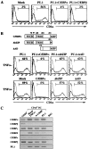
Reciprocal roles for C/EBP and PU.1 in the development of LCs. (A) The inhibition of PU.1-dependent LC differentiation by cotransduced wild-type C/EBP. CD34+ progenitors cotransduced with PU.1 and either C/EBPα or C/EBPβ were cultured for 10 d in the presence of SCF and GM-CSF. Then, CD1a expression was analyzed by flow cytometry. (B) The effects of C/EBPα mutants on DC differentiation. Schematic representation of C/EBPα mutants (upper panel). TE, transactivation element; N, nuclear localization signal. CD34+ progenitors cotransduced with PU.1 and C/EBPα mutants were cultured for 10 d in the presence of SCF and GM-CSF only (middle panel). CD34+ progenitors that only transduced with C/EBPα mutants were similarly processed in the presence of SCF, GM-CSF, and TNFα (lower panel). Then, CD1a expression was analyzed by flow cytometry. (C) RNA expression of endogenous C/EBP and PU.1 in transduced cells by RT-PCR. Cord blood CD34+ cells (CD34+) were collected by cell sorting. Cord blood CD34+ cells transduced with empty vector (mock) were cultured for 10 d in the presence of SCF, GM-CSF, and TNFα. Cord blood CD34+ cells transduced with either with A-C/EBP or PU.1 were similarly processed in the presence of SCF and GM-CSF only. Then, CD1a+ DCs were collected by cell sorting. Lane H2O represents the negative control without template.
Figure 8.
Figure 8.
Model for the role of C/EBP and PU.1 in lineage commitment. As explained in the text, C/EBP activity is essential for the commitment of pluripotent myeloid progenitors into granulocyte/macrophage lineage. With impaired C/EBP function, pluripotent myeloid progenitors take an alternative differentiation pathway of LC. On the other hand, the enforced expression of PU.1 in pluripotent myeloid progenitor cells promotes LC commitment, whereas that of C/EBP promotes granulocytic differentiation and inhibits PU.1-induced LC commitment. These findings suggest that the lineage commitment of pluripotent myeloid progenitor cells is mediated by functional balance between PU.1 and C/EBP transcription factors. Eo, eosinophils; Neu, neutrophils; Macro, macrophages.

Similar articles

Cited by

References

    1. Metcalf, D. 1989. The molecular control of cell division, differentiation commitment and maturation in haemopoietic cells. Nature. 339:27–30. - PubMed
    1. Tenen, D.G., R. Hromas, J.D. Licht, and D.E. Zhang. 1997. Transcription factors, normal myeloid development, and leukemia. Blood. 90:489–519. - PubMed
    1. Tamura, T., T. Nagamura-Inoue, Z. Shmeltzer, T. Kuwata, and K. Ozato. 2000. ICSBP directs bipotential myeloid progenitor cells to differentiate into mature macrophages. Immunity. 13:155–165. - PubMed
    1. Passegué, E., W. Jochum, M. Schorpp-Kistner, U. Möhle-Steinlein, and E.F. Wagner. 2001. Chronic myeloid leukemia with increased granulocyte progenitors in mice lacking JunB expression in the myeloid lineage. Cell. 104:21–32. - PubMed
    1. Kelly, L.M., U. Englmeier, I. Lafon, M.H. Sieweke, and T. Graf. 2000. MafB is an inducer of monocytic differentiation. EMBO J. 19:1987–1997. - PMC - PubMed

Publication types

MeSH terms

Substances