. 2002 Mar 4;195(5):617-24.
doi: 10.1084/jem.20011889.

Natural killer T cell ligand alpha-galactosylceramide enhances protective immunity induced by malaria vaccines

Affiliations

Natural killer T cell ligand alpha-galactosylceramide enhances protective immunity induced by malaria vaccines

Gloria Gonzalez-Aseguinolaza et al. J Exp Med. .

Abstract

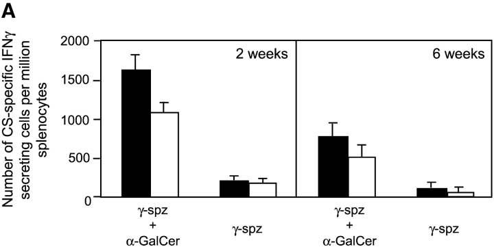
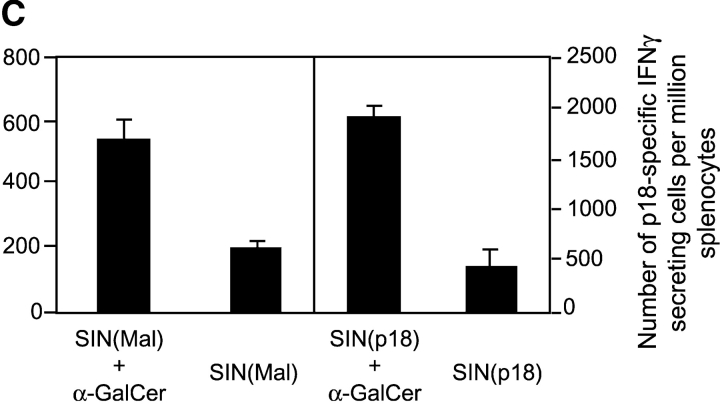
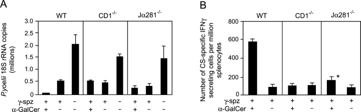
The important role played by CD8(+) T lymphocytes in the control of parasitic and viral infections, as well as tumor development, has raised the need for the development of adjuvants capable of enhancing cell-mediated immunity. It is well established that protective immunity against liver stages of malaria parasites is primarily mediated by CD8(+) T cells in mice. Activation of natural killer T (NKT) cells by the glycolipid ligand, alpha-galactosylceramide (alpha-GalCer), causes bystander activation of NK, B, CD4(+), and CD8(+) T cells. Our study shows that coadministration of alpha-GalCer with suboptimal doses of irradiated sporozoites or recombinant viruses expressing a malaria antigen greatly enhances the level of protective anti-malaria immunity in mice. We also show that coadministration of alpha-GalCer with various different immunogens strongly enhances antigen-specific CD8(+) T cell responses, and to a lesser degree, Th1-type responses. The adjuvant effects of alpha-GalCer require CD1d molecules, Valpha14 NKT cells, and interferon gamma. As alpha-GalCer stimulates both human and murine NKT cells, these findings should contribute to the design of more effective vaccines against malaria and other intracellular pathogens, as well as tumors.


Figures

Figure 1.
Figure 1.
α-GalCer enhances protective anti-malaria immunity induced by irradiated sporozoites and recombinant viruses expressing a plasmodial antigen. (A) Groups of BALB/c mice were coinjected intraperitoneally with different doses of α-GalCer (0.5, 1, or 2 μg) or vehicle (−), together with intravenous immunization with P. yoelii irradiated sporozoites (γ-spz). 2 wk later all groups of mice were challenged with infective sporozoites, and the amount of parasite ribosomal RNA in the livers was measured by real-time PCR. Sera from immunized and nonimmunized mice were collected and their titers of anti-sporozoite antibodies assayed by IFA. (B) A single dose of α-GalCer was administered 2 d before (−2), the same day (0) or 2 d after (+2) immunization with γ-spz into BALB/c (black bars) or B10.D2 (white bars) mice. (C) A group of BALB/c mice was immunized subcutaneously with a recombinant adenovirus expressing the P. yoelii CS protein, AdPyCS, together with subcutaneous administration of α-GalCer (+) or vehicle (−). (D) A group of BALB/c mice was immunized subcutaneously with a recombinant Sindbis virus expressing a CD8+ T cell epitope of the P. yoelii CS protein, SIN(Mal), together with subcutaneous administration of α-GalCer (+) or vehicle (−). Asterisk (*) indicates a significant (P < 0.01) difference between the two values using an unpaired t test. In B–D, all groups of mice were infected with live P. yoelii sporozoites 2 wk later, and the parasite burden in the liver was determined, as described in panel A. Results are expressed as the mean values ± SD of five mice.
Figure 1.
Figure 1.
α-GalCer enhances protective anti-malaria immunity induced by irradiated sporozoites and recombinant viruses expressing a plasmodial antigen. (A) Groups of BALB/c mice were coinjected intraperitoneally with different doses of α-GalCer (0.5, 1, or 2 μg) or vehicle (−), together with intravenous immunization with P. yoelii irradiated sporozoites (γ-spz). 2 wk later all groups of mice were challenged with infective sporozoites, and the amount of parasite ribosomal RNA in the livers was measured by real-time PCR. Sera from immunized and nonimmunized mice were collected and their titers of anti-sporozoite antibodies assayed by IFA. (B) A single dose of α-GalCer was administered 2 d before (−2), the same day (0) or 2 d after (+2) immunization with γ-spz into BALB/c (black bars) or B10.D2 (white bars) mice. (C) A group of BALB/c mice was immunized subcutaneously with a recombinant adenovirus expressing the P. yoelii CS protein, AdPyCS, together with subcutaneous administration of α-GalCer (+) or vehicle (−). (D) A group of BALB/c mice was immunized subcutaneously with a recombinant Sindbis virus expressing a CD8+ T cell epitope of the P. yoelii CS protein, SIN(Mal), together with subcutaneous administration of α-GalCer (+) or vehicle (−). Asterisk (*) indicates a significant (P < 0.01) difference between the two values using an unpaired t test. In B–D, all groups of mice were infected with live P. yoelii sporozoites 2 wk later, and the parasite burden in the liver was determined, as described in panel A. Results are expressed as the mean values ± SD of five mice.
Figure 2.
Figure 2.
α-GalCer increases the level of antigen-specific T cell responses elicited by various vaccines. (A) A group of BALB/c mice was immunized subcutaneously with γ-spz together with or without administration of α-GalCer by the same route, and 2 or 6 wk later splenic lymphocytes were isolated and the number of IFN-γ–secreting CS-specific CD8+ (black bars) and CD4+ (white) T cells was determined by an ELISPOT assay. (B) A group of BALB/c mice was immunized subcutaneously with AdPyCS together with or without subcutaneous administration of α-GalCer (+) or vehicle (−). 2 wk later the number of IFN-γ secreting CS-specific CD8+ (black bars) and CD4+ (white bars) T cells was determined by an ELISPOT assay. (C) A group of BALB/c mice was immunized subcutaneously with SIN(Mal) or SIN(p18) together with or without subcutaneous administration of α-GalCer. 2 wk later the number of IFN-γ secreting CS-specific and p18-specific CD8+ T cells was determined by an ELISPOT assay. The data represent one of two experiments with similar results and are expressed as the mean values ± SD of three mice.
Figure 2.
Figure 2.
α-GalCer increases the level of antigen-specific T cell responses elicited by various vaccines. (A) A group of BALB/c mice was immunized subcutaneously with γ-spz together with or without administration of α-GalCer by the same route, and 2 or 6 wk later splenic lymphocytes were isolated and the number of IFN-γ–secreting CS-specific CD8+ (black bars) and CD4+ (white) T cells was determined by an ELISPOT assay. (B) A group of BALB/c mice was immunized subcutaneously with AdPyCS together with or without subcutaneous administration of α-GalCer (+) or vehicle (−). 2 wk later the number of IFN-γ secreting CS-specific CD8+ (black bars) and CD4+ (white bars) T cells was determined by an ELISPOT assay. (C) A group of BALB/c mice was immunized subcutaneously with SIN(Mal) or SIN(p18) together with or without subcutaneous administration of α-GalCer. 2 wk later the number of IFN-γ secreting CS-specific and p18-specific CD8+ T cells was determined by an ELISPOT assay. The data represent one of two experiments with similar results and are expressed as the mean values ± SD of three mice.
Figure 3.
Figure 3.
The adjuvant activity of α-GalCer requires CD1d molecules and Vα14 NKT cells. (A) Groups of CD1d-deficient (CD1−/−), Vα14 NKT (Jα281−/−) deficient and wild-type (WT) mice on a BALB/c background were immunized intravenously with γ-spz together with intraperitoneal administration of α-GalCer (+) or vehicle (−). 2 wk later these and nonimmunized mice were challenged with viable sporozoites, and the parasite burden in the liver was measured as described in Fig. 1. (B) Identical groups of mice as described in panel A were immunized with γ-spz with intraperitoneal injection of α-GalCer (+) or vehicle (−). 2 wk later the number of IFN-γ secreting CS-specific CD8+ T cells in the spleens was determined by an ELISPOT assay. Asterisk (*) indicates a significant (P < 0.01) difference between the two values using an unpaired t test. The results reflect two experiments with similar results and are expressed as the mean values ± SD of five (A) or three (B) mice.
Figure 4.
Figure 4.
The adjuvant activity of α-GalCer is abolished in IFN-γ receptor-deficient mice. (A) Groups of IFN-γ receptor-deficient (IFN-γ R / ) and wild-type (WT) mice on a B10.D2 background were immunized intravenously with γ-spz together with intraperitoneal administration of α-GalCer (+) or vehicle (−). 2 wk later splenic lymphocytes were obtained and the number of IFN-γ secreting CS-specific CD8+ (black bars) and CD4+ (white bars) T cells were determined by an ELISPOT assay. (B) Hepatic lymphocytes were obtained from IFN-γ R / and wild-type mice and stained with PE-labeled CD1d/α-GalCer tetramer and FITC-labeled anti-CD3 antibody, and the percentage of α-GalCer–specific T cells was determined by flow cytometric analysis. The number indicated in the upper right corners represents the percentage of double-positive cells among the liver lymphoid cell population. (C) Hepatic lymphocytes were obtained from IFN-γ R / (black bars) or WT (white bars) mice, and the number of IFN-γ or IL-4 secreting α-GalCer–specific cells were determined by an ELISPOT assay. Results are expressed as the mean values ± SD of five mice.

Comment in

References

    1. Seder, R.A., and A.V. Hill. 2000. Vaccines against intracellular infections requiring cellular immunity. Nature. 406:793–798. - PubMed
    1. Smyth, M.J., D.T. Godfrey, and J.A. Trapani. 2001. A fresh look at tumor immunosurveillance and immunotherapy. Nat. Immunol. 2:293–299. - PubMed
    1. Pardoll, D.M., and S.L. Topalian. 1998. The role of CD4+ T cell responses in antitumor immunity. Curr. Opin. Immunol. 10:588–594. - PubMed
    1. Singh, M., and D. O'Hagan. 1999. Advances in vaccine adjuvants. Nat. Biotechnol. 17:1075–1081. - PubMed
    1. de Gruijl, T.D., and D.T. Curiel. 1999. Cancer vaccine strategies get bigger and better. Nat. Med. 5:1124–1125. - PubMed

Publication types