. 2002 Mar 4;195(5):643-9.
doi: 10.1084/jem.20012028.

Specificity of tissue transglutaminase explains cereal toxicity in celiac disease

Affiliations

Specificity of tissue transglutaminase explains cereal toxicity in celiac disease

L Willemijn Vader et al. J Exp Med. .

Abstract

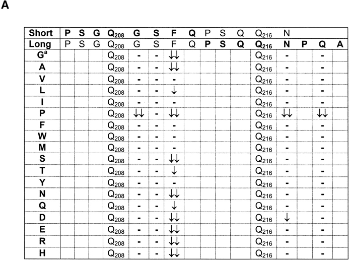
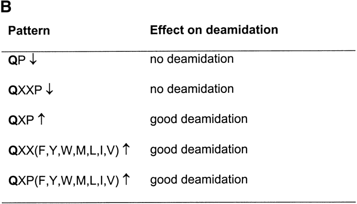
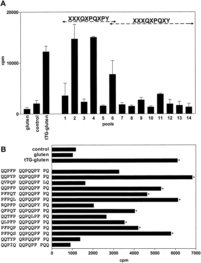
Celiac disease is caused by a selective lack of T cell tolerance for gluten. It is known that the enzyme tissue transglutaminase (tTG) is involved in the generation of T cell stimulatory gluten peptides through deamidation of glutamine, the most abundant amino acid in gluten. Only particular glutamine residues, however, are modified by tTG. Here we provide evidence that the spacing between glutamine and proline, the second most abundant amino acid in gluten, plays an essential role in the specificity of deamidation. On the basis of this, algorithms were designed and used to successfully predict novel T cell stimulatory peptides in gluten. Strikingly, these algorithms identified many similar peptides in the gluten-like hordeins from barley and secalins from rye but not in the avenins from oats. The avenins contain significantly lower percentages of proline residues, which offers a likely explanation for the lack of toxicity of oats. Thus, the unique amino acid composition of gluten and related proteins in barley and rye favors the generation of toxic T cell stimulatory gluten peptides by tTG. This provides a rationale for the observation that celiac disease patients are intolerant to these cereal proteins but not to other common food proteins.


Figures

Figure 1.
Figure 1.
The influence of amino acid substitutions on deamidation of Q208 and Q216 in an HLA-DQ8–restricted gliadin peptide. (A) Overview of the influence of COOH-terminal amino acid substitutions on deamidation of Q208 in the short version of the gliadin peptide and Q216 in the long version of the peptide. aThe amino acids G209, S210, F211, N217, and Q219 were substituted for all amino acids except lysine and cysteine, because these residues could affect deamidation by formation of disulphide bridges and tTG driven cross-linking, respectively. The effect on deamidation was determined by mass spectrometry. Substitutions that resulted in major differences in deamidation are designated with arrows: ↓↓ decrease of 70–100%; ↓ decrease of 30–69%. Dash indicates no effect of the substitution; substitution at other positions in the peptide had only minor effects on deamidation of the Q208 and Q216 residues (data not shown). (B) Patterns for deamidation in the HLA-DQ8–restricted gliadin peptide. X stands for any amino acid.
Figure 1.
Figure 1.
The influence of amino acid substitutions on deamidation of Q208 and Q216 in an HLA-DQ8–restricted gliadin peptide. (A) Overview of the influence of COOH-terminal amino acid substitutions on deamidation of Q208 in the short version of the gliadin peptide and Q216 in the long version of the peptide. aThe amino acids G209, S210, F211, N217, and Q219 were substituted for all amino acids except lysine and cysteine, because these residues could affect deamidation by formation of disulphide bridges and tTG driven cross-linking, respectively. The effect on deamidation was determined by mass spectrometry. Substitutions that resulted in major differences in deamidation are designated with arrows: ↓↓ decrease of 70–100%; ↓ decrease of 30–69%. Dash indicates no effect of the substitution; substitution at other positions in the peptide had only minor effects on deamidation of the Q208 and Q216 residues (data not shown). (B) Patterns for deamidation in the HLA-DQ8–restricted gliadin peptide. X stands for any amino acid.
Figure 2.
Figure 2.
Stimulation of gluten-specific T cell lines by predicted peptides. (A) A gluten-specific T cell line isolated from a small intestinal biopsy of a CD patient was tested against gluten, tTG-treated gluten, and against the predicted peptides that were present in pools of five peptides each (pools 1–14). The odd numbers represent pools that contain the native peptides, whereas the even numbers represent corresponding pools that were treated with tTG. Strong reactivity of the T cell line was only observed with tTG-treated gluten and tTG-treated pools of peptides predicted by the XXXQXPQXPY algorithm. Cpm indicates 3[H]thymidine incorporation. (B) Reactivity of the T cell line against the individual peptides from the T cell stimulatory pools. Bars indicated with an asterisk are considered positive (Stimulation Index > 3).

Similar articles

Cited by

References

    1. Marsh, M.N. 1992. Gluten, major histocompatibility complex, and the small intestine. A molecular and immunobiologic approach to the spectrum of gluten sensitivity (‘celiac sprue’). Gastroenterology. 102:330–354. - PubMed
    1. Dieterich, W., T. Ehnis, M. Bauer, P. Donner, U. Volta, E.O. Riecken, and D. Schuppan. 1997. Identification of tissue transglutaminase as the autoantigen of celiac disease. Nat. Med. 3:797–801. - PubMed
    1. Molberg, O., S.N. McAdam, R. Korner, H. Quarsten, C. Kristiansen, L. Madsen, L. Fugger, H. Scott, O. Noren, P. Roepstorff, K.E. Lundin, H. Sjostrom, and L.M. Sollid. 1998. Tissue transglutaminase selectively modifies gliadin peptides that are recognized by gut-derived T cells in celiac disease. Nat. Med. 4:713–717. - PubMed
    1. van de Wal, Y., Y. Kooy, P. van Veelen, S. Pena, L. Mearin, G. Papadopoulos, and F. Koning. 1998. Selective deamidation by tissue transglutaminase strongly enhances gliadin-specific T cell reactivity. J. Immunol. 161:1585–1588. - PubMed
    1. Spurkland, A., G. Ingvarsson, E.S. Falk, I. Knutsen, L.M. Sollid, and E. Thorsby. 1997. Dermatitis herpetiformis and celiac disease are both primarily associated with the HLA-DQ (α1*0501, β1*02) or the HLA-DQ (α1*03, β1*0302) heterodimers. Tiss. Anttigens. 49:29–34. - PubMed

Publication types