. 2002 Mar 4;195(5):651-6.
doi: 10.1084/jem.20012023.

Rapid cytotoxic T lymphocyte activation occurs in the draining lymph nodes after cutaneous herpes simplex virus infection as a result of early antigen presentation and not the presence of virus

Affiliations

Rapid cytotoxic T lymphocyte activation occurs in the draining lymph nodes after cutaneous herpes simplex virus infection as a result of early antigen presentation and not the presence of virus

Scott N Mueller et al. J Exp Med. .

Abstract

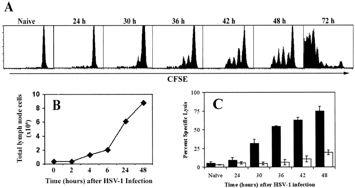
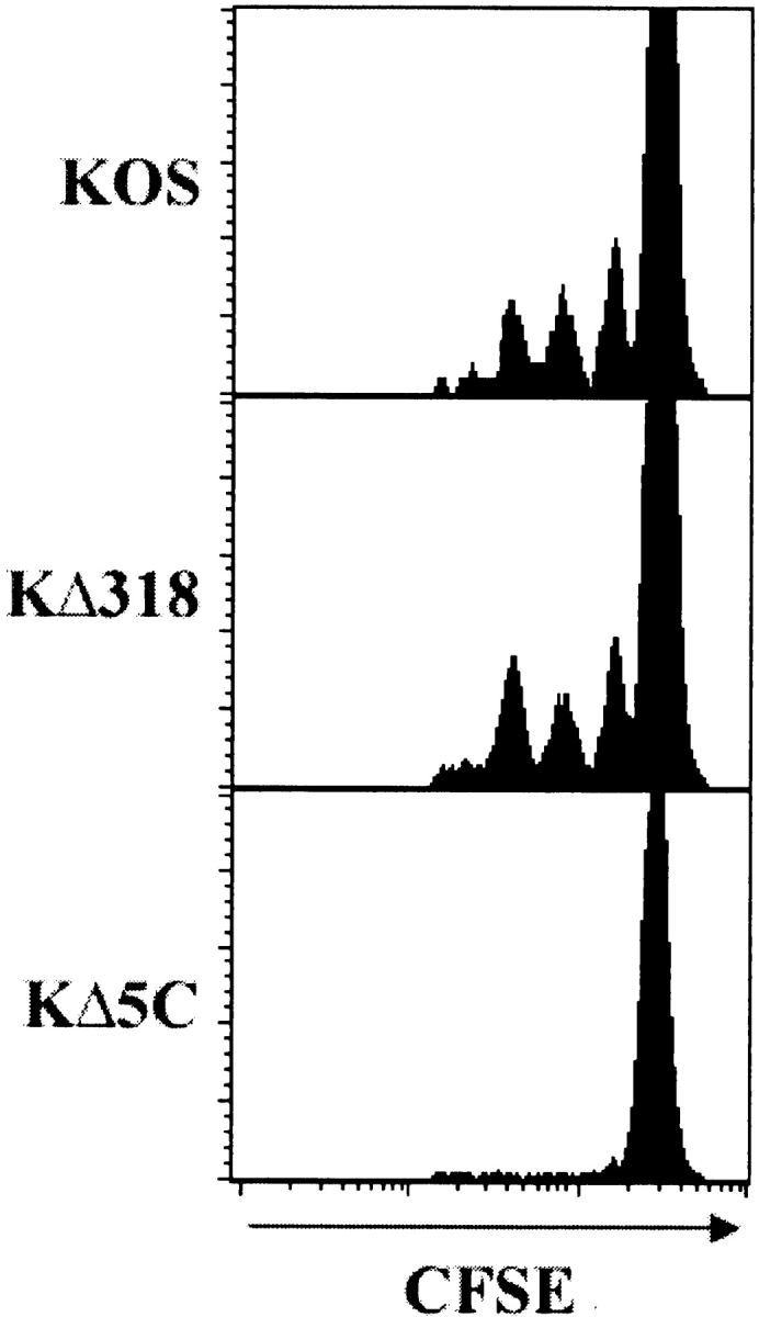
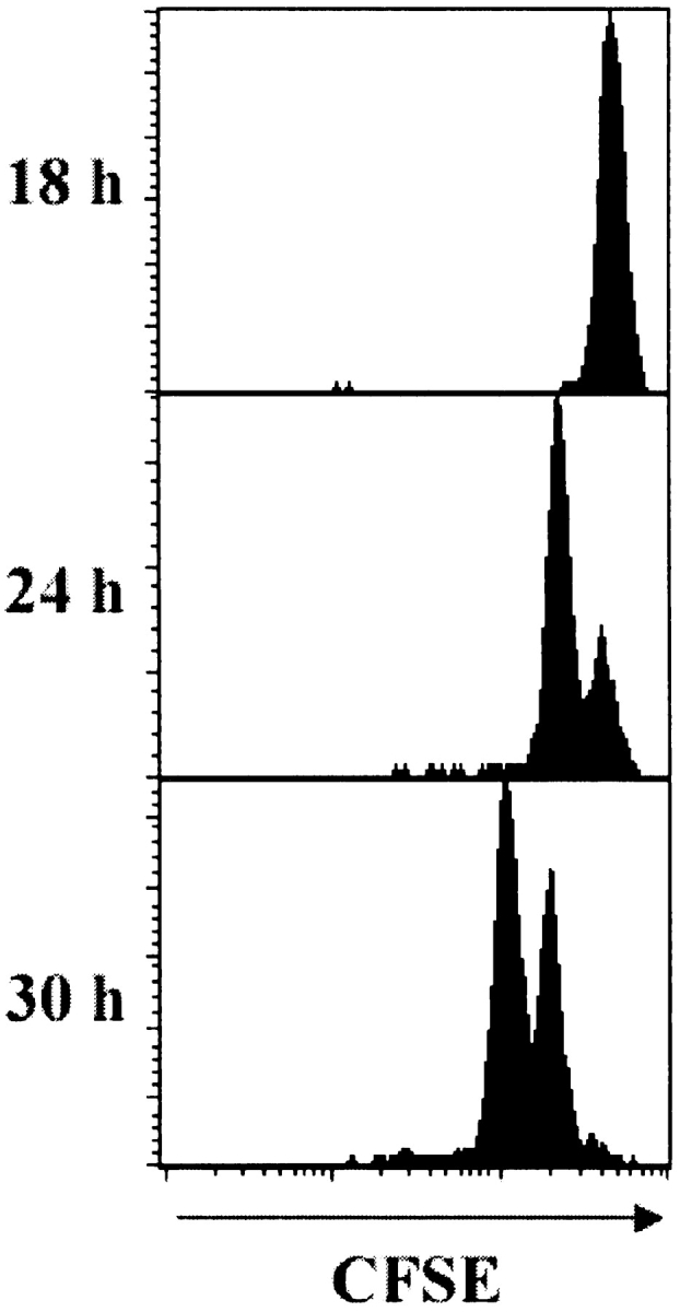
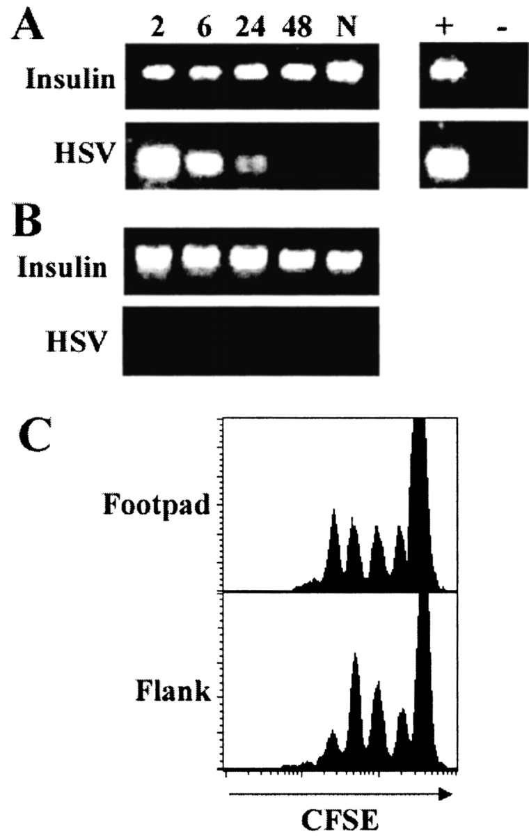
Localized cutaneous herpes simplex virus type 1 (HSV-1) infection leads to arming and initial expansion of cytotoxic T lymphocytes (CTLs) in the draining popliteal lymph nodes (PLNs) followed by migration and further proliferation in the spleen. To accurately characterize the sequence of events involved in the activation and generation of anti-HSV CTLs, we used T cell receptor (TCR) transgenic mice specific for the immunodominant epitope from HSV glycoprotein B (gB(498-505)). We describe the detection of the initiation of antigen presentation in the draining lymph nodes by 4-6 h after infection with HSV-1. Analysis of CD69 up-regulation revealed activation of gB-specific CD8(+) T cells by 6-8 h after infection. Furthermore, we show that T cell proliferation begins no sooner than 24 h after activation and is marked by the concurrent appearance of CTL activity in the PLNs. These events are not dependent on the presence of virus in the draining lymph nodes, and suggest a requirement for recruitment of professional antigen-presenting cells to the site of T cell activation. Consequently, we have defined the initiation of the CD8(+) T cell-mediated response to cutaneous HSV-1 infection, demonstrating that the immune response to localized viral infection depends only on the appearance of cells presenting virus-derived antigen and commences with remarkable swiftness.


Figures

Figure 1.
Figure 1.
Concurrent in vivo proliferation and CTL activity by gB-specific CD8+ T cells in the PLNs after cutaneous infection with HSV-1. (A) CFSE-labeled lymph node cells from gBT-I.1 mice were transferred into C57BL/6 mice before infection with HSV-1. PLN cells were isolated at various times after infection (24–72 h) and dilution of the CFSE fluorescence analyzed by gating on live CD8+ T cells. (B) Cellularity within the draining lymph nodes over a 48-h period was determined using cell suspensions obtained from the PLNs of mice after foot-pad HSV-1 infection. (C) Mice that had (black bars) or had not (white bars) received 106 gBT-I.1 cells 24 h earlier were infected with HSV-1 in the footpad and left for various times as shown before intravenous transfer of CFSE-labeled syngeneic target cells. gB-peptide–pulsed splenocytes were labeled with a high concentration of CFSE (CFSEhi) while unpulsed control targets were labeled with a low concentration of CFSE (CFSElo). 4 h after target cell transfer, mice were killed and PLN cells analyzed for relative elimination of the CFSEhi versus CFSElo populations. Percent specific lysis was calculated as described in reference 5. Error bars represent SD.
Figure 2.
Figure 2.
De novo synthesis of viral peptides is required to elicit a gB-specific T cell response. Mice receiving CFSE-labeled gBT-I.1 CD8+ T cells were infected with wild-type HSV-1 KOS or the gB mutant strains KΔ318 or KΔ5C. 42 h after infection, CD8+ T cells from draining PLNs were analyzed for dilution of the CFSE stain caused by cell division. Histograms represent 5–10,000 live events.
Figure 3.
Figure 3.
Naive T cells require a minimum 24-h lag between activation and commencement of proliferation. Mice receiving CFSE-labeled gBT-I.1 cells were immunized intravenously with gB498–505 peptide and the transgenic CD8+ T cells in the spleen analyzed 18, 24, and 30 h later for the presence of dividing cells. Histograms represent 5–10,000 live events.
Figure 4.
Figure 4.
Activation of CD8+ T cells in the PLNs correlates with the appearance of specific antigen presentation. (A) Mice adoptively transferred with CFSE-labeled gBT-I.1 cells were killed at various times (2–8 h) after footpad infection with HSV-1 and CD8+CFSE+ cells analyzed for the expression of CD69. (B) PLNs from HSV-1–infected C57BL/6 mice were treated with collagenase to form a cell suspension. Graded amounts of these cells were placed into culture with HSV-2.3.2E2 lacZ-inducible hybridoma cells overnight. An X-Gal assay was then performed to stain responding hybridoma cells which were counted microscopically. Numbers presented represent the total number of lacZ + cells per separate PLN at various times (2–48 h) after infection, and error bars represent SD (n = 8–12).
Figure 5.
Figure 5.
Activation of the gB-specific T cells does not require the presence of viral DNA in the draining lymph nodes. Mice were killed at various times (2–48 h; N, naive) after footpad infection (A), or after flank scarification (B) and DNA isolated from the draining PLNs or pooled axillary and inguinal lymph nodes, respectively. 100 ng of DNA was amplified by PCR using HSV-1 or insulin-specific primers. DNA from the footpad of a mouse infected 24 h earlier was used as a positive control (+). A no DNA control was included to rule out contamination (−). (C) Mice receiving CFSE-labeled gBT-I.1 cells were infected with HSV-1 by footpad injection or flank scarification, and CD8+ T cells from the PLNs or pooled axillary and inguinal lymph nodes, respectively, were analyzed for the presence of proliferating cells via CFSE intensity at 48 h after infection. Histograms represent 5–10,000 live events.

Similar articles

Cited by

References

    1. Banchereau, J., and R.M. Steinman. 1998. Dendritic cells and the control of immunity. Nature. 392:245–252. - PubMed
    1. Macatonia, S.E., S.C. Knight, A.J. Edwards, S. Griffiths, and P. Fryer. 1987. Localization of antigen on lymph node dendritic cells after exposure to the contact sensitizer fluorescein isothiocyanate. Functional and morphological studies. J. Exp. Med. 166:1654–1667. - PMC - PubMed
    1. Cose, S.C., C.M. Jones, M.E. Wallace, W.R. Heath, and F.R. Carbone. 1997. Antigen-specific CD8+ T cell subset distribution in lymph nodes draining the site of herpes simplex virus infection. Eur. J. Immunol. 27:2310–2316. - PubMed
    1. McNally, J.M., D. Dempsey, R.M. Wolcott, R. Chervenak, and S.R. Jennings. 1999. Phenotypic identification of antigen-dependent and antigen-independent CD8 CTL precursors in the draining lymph node during acute cutaneous herpes simplex virus type 1 infection. J. Immunol. 163:675–681. - PubMed
    1. Coles, R.M., S.N. Mueller, W.R. Heath, F.R. Carbone, and A.G. Brooks. 2002. Progression of armed CTL from draining lymph node to spleen shortly after localised infection with HSV-1. J. Immunol. 168:834–838. - PubMed

Publication types

Substances