. 2002 Mar;2(3):319-30.
doi: 10.1016/s1534-5807(02)00135-1.

Conserved organization of centromeric chromatin in flies and humans

Affiliations

Conserved organization of centromeric chromatin in flies and humans

Michael D Blower et al. Dev Cell. 2002 Mar.

Abstract

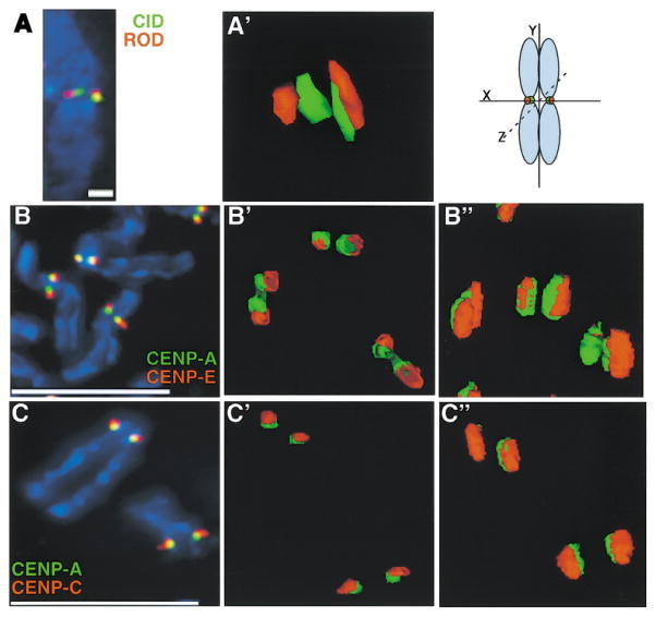
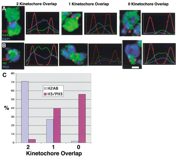
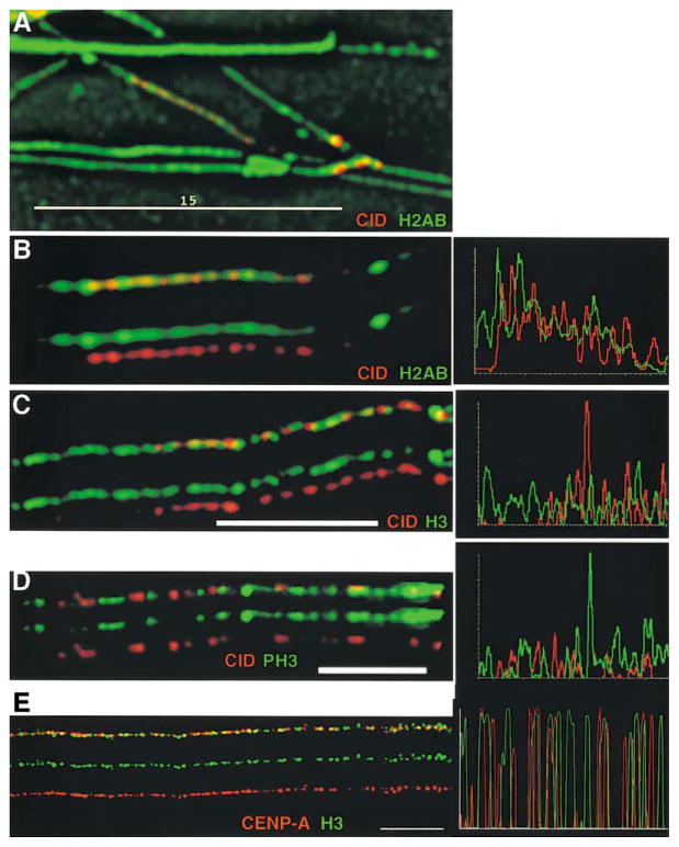
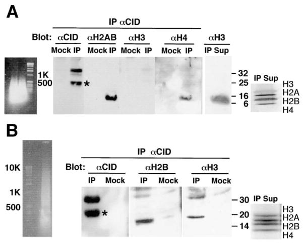
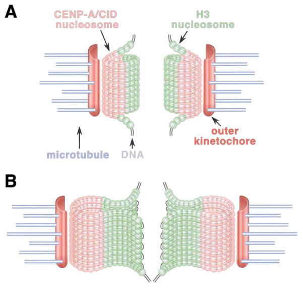
Recent studies have highlighted the importance of centromere-specific histone H3-like (CENP-A) proteins in centromere function. We show that Drosophila CID and human CENP-A appear at metaphase as a three-dimensional structure that lacks histone H3. However, blocks of CID/CENP-A and H3 nucleosomes are linearly interspersed on extended chromatin fibers, and CID is close to H3 nucleosomes in polynucleosomal preparations. When CID is depleted by RNAi, it is replaced by H3, demonstrating flexibility of centromeric chromatin organization. Finally, contrary to models proposing that H3 and CID/CENP-A nucleosomes are replicated at different times in S phase, we show that interspersed H3 and CID/CENP-A chromatin are replicated concurrently during S phase in humans and flies. We propose that the unique structural arrangement of CID/CENP-A and H3 nucleosomes presents centromeric chromatin to the poleward face of the condensing mitotic chromosome.


Figures

Figure 1
Figure 1. CENP-A Proteins form a Higher Order Structure on Metaphase Chromosomes in both Drosophila and Humans
(A) CID (green) and ROD (Rough Deal) (red) were colocalized on three-dimensional meta-phase chromosomes from Drosophila S2 cells and showed typical double-dot staining by immunofluorescence and subsequent modeling of antibody staining. (A′) Rotation of the chromosome 90° around the x axis showed that the double-dot staining of CID appears as a cylindrical structure that spans the depth of the chromosome, and that ROD is located next to, but minimally overlaps with, the CID cylinder. Scale bar is 5 microns. (B and B′) CENP-E (red), a mammalian outer kinetochore protein, shows minimal overlap with CENP-A (green) by deconvolution microscopy. (B″) Rotation of the chromosome by 35° around the x axis showed a similar cylindrical structure for CENP-A as that observed for CID at Drosophila kinetochores. Scale bar is 15 microns. (C and C′) CENP-C (red), an inner kinetochore protein, typically shows more overlap with CENP-A (green) by 2D immunofluorescence than does CENP-E. (C″) Rotation by 70° around the x axis showed that CENP-C also appears to be wrapped around the CENP-A structure and does not overlap significantly with CENP-A. Scale bar 15 microns. See text and Experimental Procedures for quantitation of the extent of overlap.
Figure 2
Figure 2. Metaphase Centromeres Contain CID and Histones H2A, H2B but Not Histone H3
CID and core histones H2A, H2B, and H3 were localized by indirect immunofluorescence on metaphase chromosomes from S2 and Kc cells, and the fluorescence intensity was quantitated across the two sister kinetochores. (A) CID shows three patterns of localization with respect to H2AB: colocalization with both kinetochores, colocalization with one kinetochore, or colocalization with neither kinetochore. (B) CID shows three patterns of localization with respect to H3/PH3: colocalization with both kinetochores, colocalization with one kinetochore, or colocalization with neither kinetochore. (C) Quantitation of the relative distribution patterns of CID localization with respect to H2AB and H3/PH3. Scale bar is 1 micron.
Figure 3
Figure 3. CID Is Colocalized with Histone H2AB on Extended Chromatin Fibers, but Is Interspersed with Histone H3
Extended chromatin fibers were prepared from Drosophila Kc and human tissue culture cells, and CID/CENP-A and other core histones were simultaneously localized by indirect immunofluorescence. Single-color images are shown under the merged images in (B)–(D). Line graph to the right of each image shows quantitation of antibody staining along the length of a fiber. (A) View of an entire field of chromatin fibers immunostained for H2AB (green) and CID (red). Most fibers show strong staining of H2AB antibodies continuously along their lengths, including the CID subdomains. Scale bar is 15 microns. (B) An isolated chromatin fiber from (A) on which CID appears punctate and discontinuous, but H2AB are continuous along the fiber. (C) CID (red) and histone H3 (green) are present in close proximity along the same chromatin fiber and appear to be interspersed with one another, with very little overlap. Scale bar is 10 microns. (D) CID (red) and histone PH3 (green) are present in close proximity along the same chromatin fiber and appear to be interspersed with one another, with some overlap. Scale bar is 5 microns. (E) Human CENP-A (green) and H3 (red) are interspersed with infrequent overlap, similar to the organization observed in Drosophila centromeric chromatin. Scale bar is 15 microns for (A), 5 microns for (D), and 10 microns for (C) and (E). See text and Experimental Procedures for quantitation of the extent of overlap.
Figure 4
Figure 4. CID Mononucleosomes Do Not Contain Histone H3, while CID Polynucleosomes Contain Histone H3
Soluble chromatin was prepared from Kc cells using micrococcal nuclease digestion, and CID-containing chromatin was immunoprecipitated using affinity-purified anti-CID. (A) Short polynucleosomes (primarily mono-nucleosomes) were prepared using extensive micrococcal nuclease digestion. CID and a degradation product (asterisk) were precipitated, as well as histones H2A, H2B and histone H4, while histone H3 was not coimmunoprecipitated from short CID-containing chromatin. Mock IPs did not result in the recovery of any histones. Histone H3 was found in the supernatant of the IP (IP Sup), and a silver-stained gel shows the integrity of the histones in the chromatin used in the IPs (note that after silver staining, H3 stains somewhat negatively compared to other core histones). (B) Polynucleosomes ranging from ~150 bp to 10 kb were prepared using limited micrococcal nuclease digestion. CID and a degradation product (asterisk) were immunoprecipitated in IPs, but not in mock IPs. Histone H2B as well as histone H3 was coimmunoprecipitated from long chromatin containing CID. Coomassie-stained gel shows the integrity of the histones in the chromatin used in the IPs.
Figure 5
Figure 5. H3 Replaces CID When CID Is Depleted by RNAi, and Centromeric Chromatin Replicates Synchronously in Human and Drosophila Cells
Single-color images are shown beneath merged images. Line graph to the right of each image shows quantitation of antibody staining along a fiber. (A) Histone H3 replaces CID when CID is depleted by RNAi. In fibers from CID RNAi-treated cells, decreased numbers of CID spots (red) are spaced further apart, and larger regions of H3 (green) are found between CID spots. Scale bar is 5 microns. (B) Replication timing of centromeric chromatin, which contains interspersed H3 and CID nucleosomes, occurs simultaneously throughout the domain. CldU incorporation (green) on fibers shows continuous staining throughout the kinetochore chromatin (identified by CID antibodies [red]), as denoted by quantitation of signal in bar graph (right). Yellow arrows denote chromatin that contains CID and CldU. Green arrows indicate regions that lack CID staining but contain CldU staining (green), indicating that H3 chromatin adjacent to blocks of CID is replicated simultaneously with CID chromatin. Scale bar is 10 microns. (C) Interspersed human CENP-A and H3 are replicated concurrently. Detection of replication by CldU incorporation (green) shows that both CENP-A (red) and the spaces between CENP-A staining which represent H3 (see Figure 2B) colocalize with CldU. Yellow arrows denote CENP-A chromatin that is replicated; green arrows indicate nearby regions that are devoid of CENP-A staining but show CldU staining (green). Arrowhead demonstrates that CldU staining abruptly ends at the edge of CENP-A staining and does not extend into the adjacent chromatin. Scale bar is 15 microns.
Figure 6
Figure 6. Models for 2D and 3D Organization of Centromeric Chromatin in Drosophila and Humans
Two models are proposed to reconcile the 2D interspersion of CENP-A and CID in centromeric chromatin fibers with the sequestration observed in three-dimensional analyses of metaphase chromosomes. (A) Solenoid model. Blocks of CENP-A/CID and H3 nucleosomes may be arranged in a solenoid, such that CID nucleosomes are present on the poleward face of the meta-phase chromosome while H3 nucleosomes are sequestered in the region between the paired sister kinetochores. In this model, the DNA is proposed to spiral through the three dimensional cylindrical structure. (B) Looping model. It is also possible that centromeric DNA may loop, rather than spiral, through the elliptical structure, resulting in CENP-A domains on the poleward face of the chromosome, and H3 domains located toward the region between sister centromeres.

References

    1. Ahmad K, Henikoff S. Centromeres are specialized replication domains in heterochromatin. J Cell Biol. 2001;153:101–109. - PMC - PubMed
    1. Bernard P, Maure JF, Partridge JF, Genier S, Javerzat JP, Allshire RC. Requirement of heterochromatin for cohesion at centromeres. Science. 2001;294:2539–2542. - PubMed
    1. Blat Y, Kleckner N. Cohesins bind to preferential sites along yeast chromosome III, with differential regulation along arms versus the centric region. Cell. 1999;98:249–259. - PubMed
    1. Blower MD, Karpen GH. The role of Drosophila CID in kinetochore formation, cell-cycle progression and heterochromatin interactions. Nat Cell Biol. 2001;3:730–739. - PMC - PubMed
    1. Buchwitz BJ, Ahmad K, Moore LL, Roth MB, Henikoff S. A histone-H3-like protein in C. elegans. Nature. 1999;401:547–548. - PubMed

Publication types

MeSH terms