. 2002 Mar 1;22(5):1618-28.
doi: 10.1523/JNEUROSCI.22-05-01618.2002.

Physiological role of calcium-activated potassium currents in the rat lateral amygdala

Affiliations

Physiological role of calcium-activated potassium currents in the rat lateral amygdala

E S Louise Faber et al. J Neurosci. .

Abstract

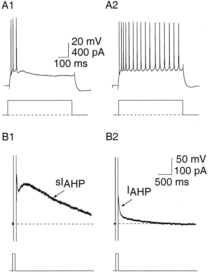
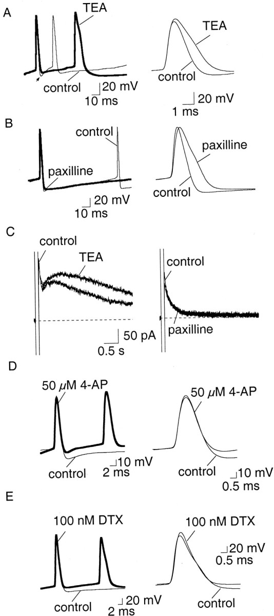
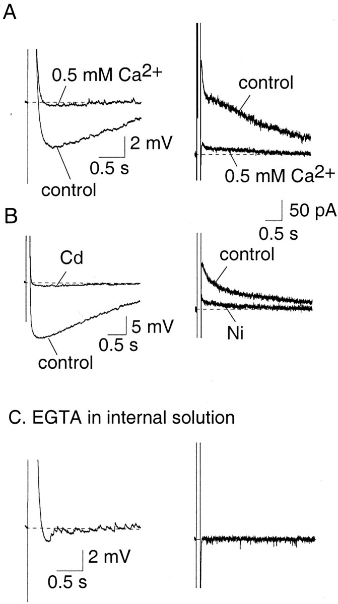
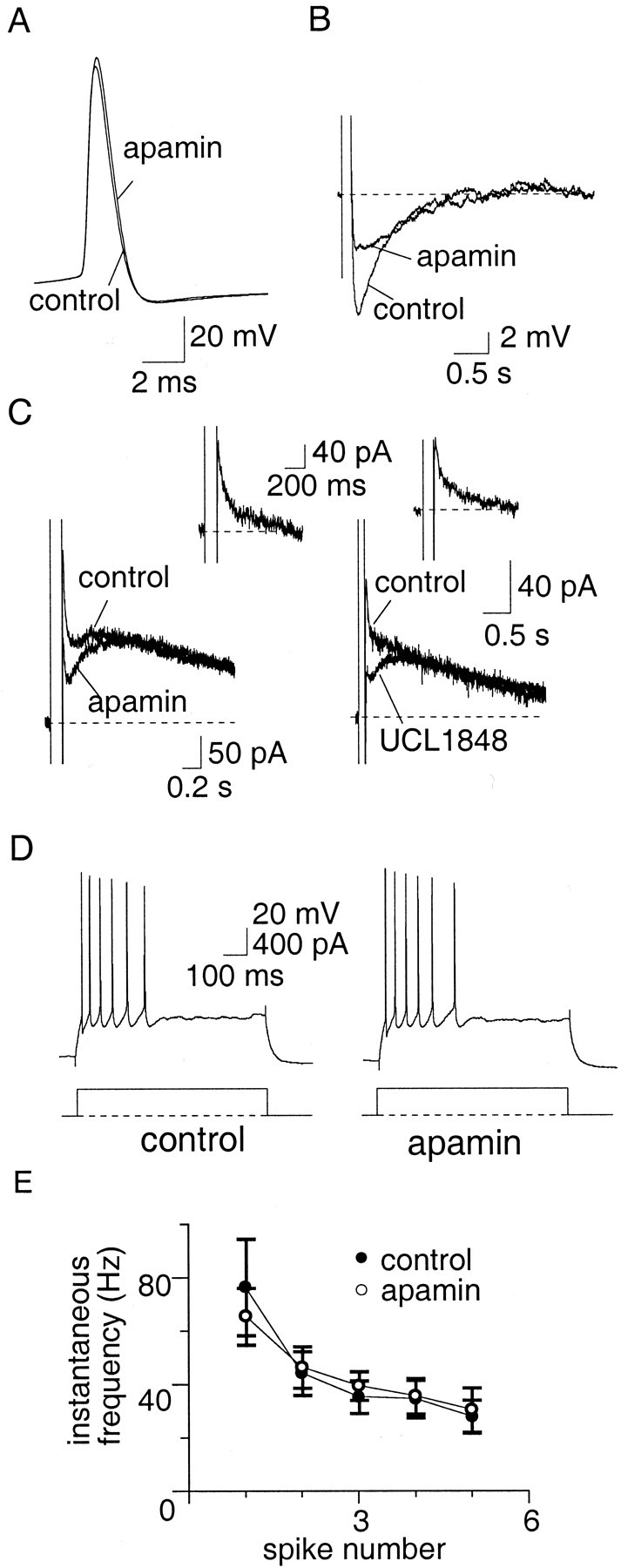
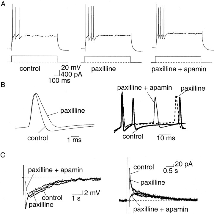
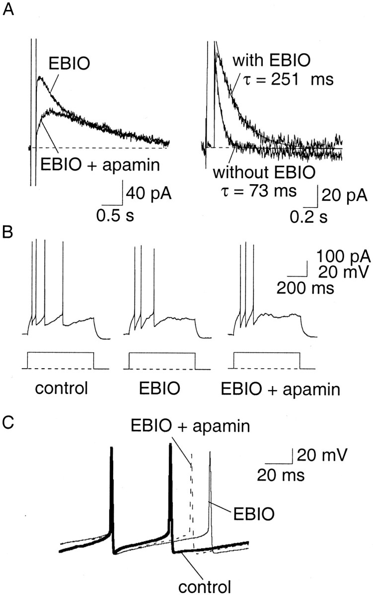
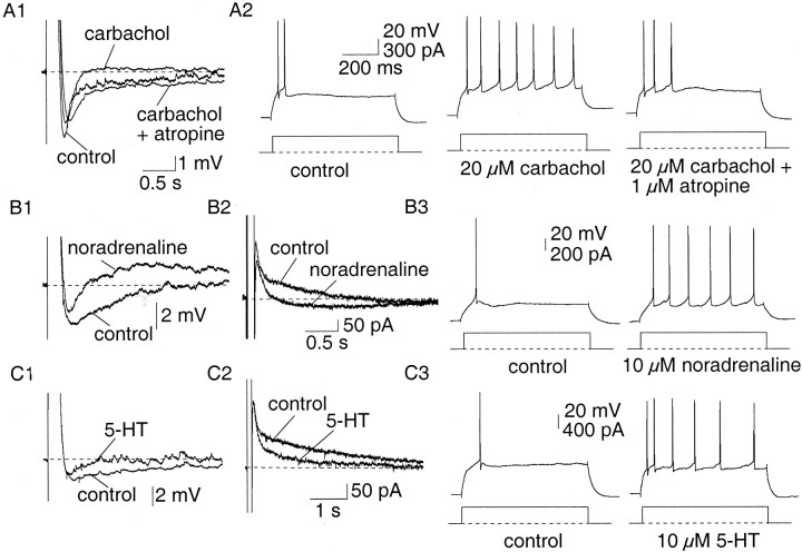
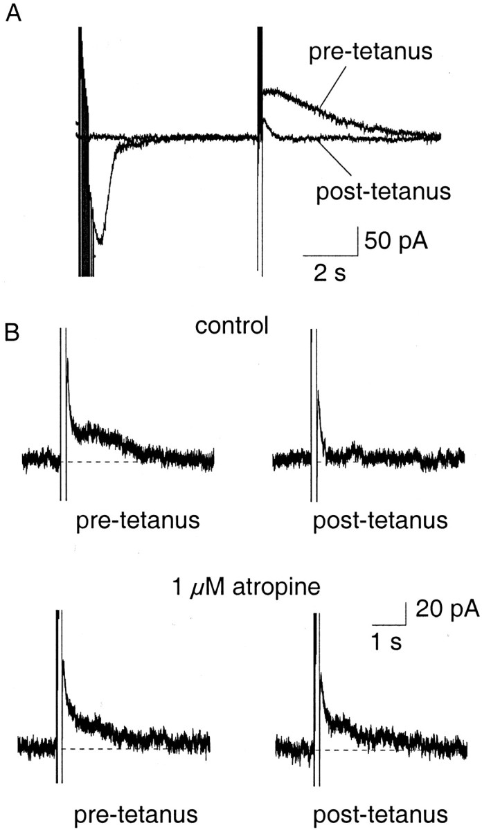
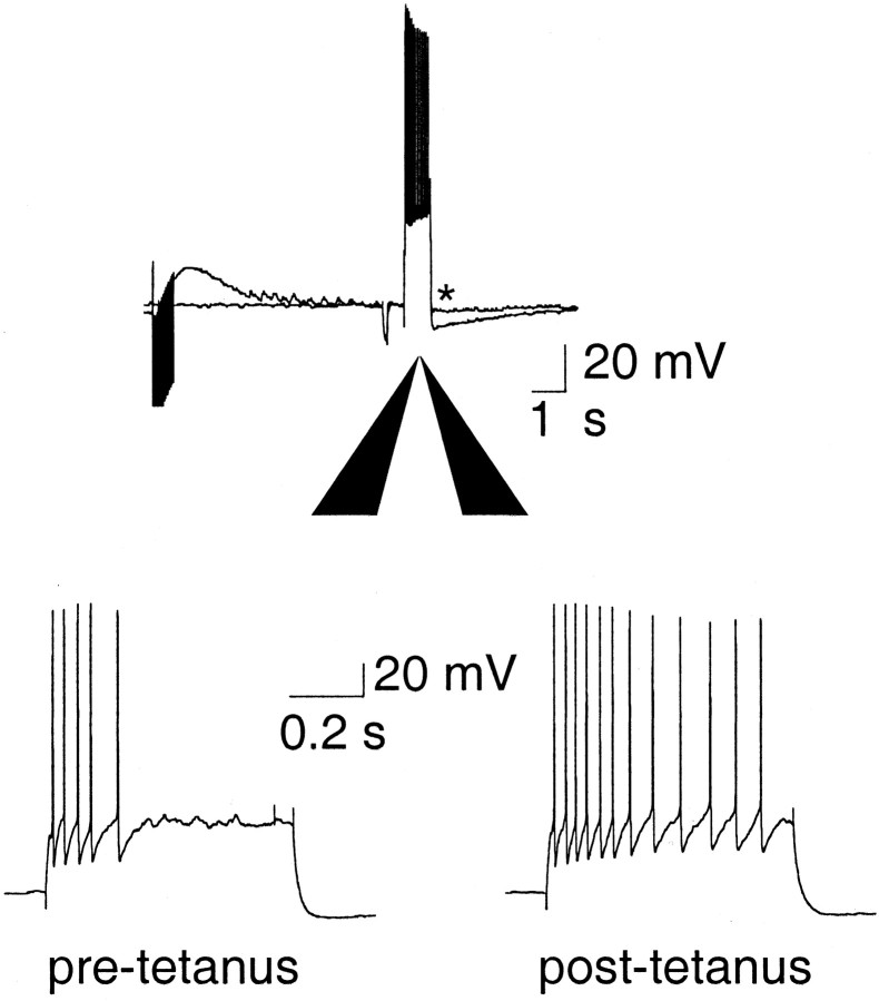
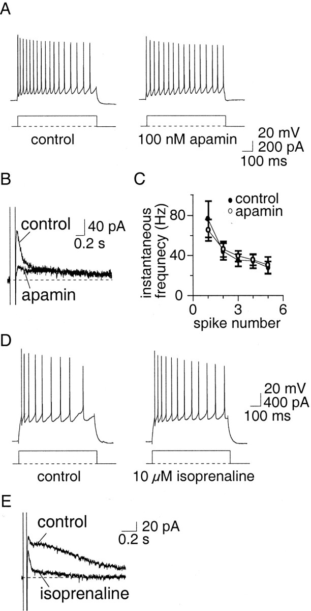
Principal neurons in the lateral nucleus of the amygdala (LA) exhibit a continuum of firing properties in response to prolonged current injections ranging from those that accommodate fully to those that fire repetitively. In most cells, trains of action potentials are followed by a slow afterhyperpolarization (AHP) lasting several seconds. Reducing calcium influx either by lowering concentrations of extracellular calcium or by applying nickel abolished the AHP, confirming it is mediated by calcium influx. Blockade of large conductance calcium-activated potassium channel (BK) channels with paxilline, iberiotoxin, or TEA revealed that BK channels are involved in action potential repolarization but only make a small contribution to the fast AHP that follows action potentials. The fast AHP was, however, markedly reduced by low concentrations of 4-aminopyridine and alpha-dendrotoxin, indicating the involvement of voltage-gated potassium channels in the fast AHP. The medium AHP was blocked by apamin and UCL1848, indicating it was mediated by small conductance calcium-activated potassium channel (SK) channels. Blockade of these channels had no effect on instantaneous firing. However, enhancement of the SK-mediated current by 1-ethyl-2-benzimidazolinone or paxilline increased the early interspike interval, showing that under physiological conditions activation of SK channels is insufficient to control firing frequency. The slow AHP, mediated by non-SK BK channels, was apamin-insensitive but was modulated by carbachol and noradrenaline. Tetanic stimulation of cholinergic afferents to the LA depressed the slow AHP and led to an increase in firing. These results show that BK, SK, and non-BK SK-mediated calcium-activated potassium currents are present in principal LA neurons and play distinct physiological roles.


Figures

Fig. 1.
Fig. 1.
Electrophysiological properties of principal neurons in the LA. A, Neurons in the LA show a continuum of firing patterns ranging from full accommodation where they fire only one to five spikes (A1) to firing repetitively (A2) in response to a 400 pA, 600 msec current injection. B, The current underlying the AHP is evoked by a 100 msec, 50 mV step and can be separated into two components,IAHP and sIAHP. Cells that accommodate more have a larger current underlying the slow AHP than those that fire repetitively. B1 shows the current evoked in the cell shown in A1, and B2 shows the current evoked in the cell shown in A2.
Fig. 2.
Fig. 2.
Currents mediated by BK channels are involved in action potential repolarization in LA neurons, but contribute little to the fast AHP. A, B, The effect of BK channel blockers TEA (1 mm, A) and paxilline (10 μm, B) on action potentials. The fast AHP is indicated by the arrow. TEA both broadens the spike (p < 0.001) and reduces the fast AHP immediately after action potentials (p < 0.005). Paxilline has little effect on the fast AHP, but significantly (p < 0.005) increased the spike half-width (B). C, BK channels do not contribute to the slow AHP. Neither TEA (1 mm,left) nor paxilline (10 μm, right) blocked the currents underlying the slow AHP. The increase in the amplitude of sIAHP in the presence of TEA is attributable to the large broadening of action poten tials and consequent increased calcium influx.D, E, Two blockers of voltage-dependent potassium currents 4-AP (C) and α-dendrotoxin (DTX, D) reduce the fast AHP that follows action potentials without slowing action potential repolarization.
Fig. 3.
Fig. 3.
Activation of the AHP current requires calcium influx and a rise in cytosolic calcium. A, The AHP recorded under current clamp (left) and the underlying current recorded in voltage clamp (right) were blocked by perfusing slices with aCSF containing 0.5 mmCa2+. B, Blocking voltage-gated calcium channels blocks the AHP (left traces, cadmium 0.25 mm) and the underlying current (right traces, nickel 5 mm). C, Inclusion of high concentrations of the calcium buffer EGTA (10 mm) in the pipette solution abolished the AHP.
Fig. 4.
Fig. 4.
Apamin-sensitive channels mediate the medium AHP but play no role in spike-frequency adaptation. A,Action potentials recorded in control aCSF and in the presence of apamin (100 nm). Apamin had no effect (p > 0.05) on the spike half-width.B, The AHP evoked by a 100 msec depolarizing current injection. In the presence of apamin, the medium AHP is blocked.C, Left, Apamin blocksIAHP but has no effect on the sIAHP. C, Right, UCL1848 also blocks theIAHP. The SK-mediatedIAHP obtained by subtraction is shown in the insets. D, Train of action potentials evoked by a 600 msec, 400 pA current injection. Apamin had no effect on spike-frequency adaptation. The instantaneous firing frequency during the spike train, and the lack of effect by apamin, is shown in E.
Fig. 5.
Fig. 5.
Enhancement of the apamin-sensitive potassium current increases the early interspike interval. Paxilline (10 μm) had no effect on spike-frequency adaptation (A) but increased the early interspike interval (B, right) by increasing the spike half-width (B, left traces), mediated by enhanced calcium influx during the spike. This effect on the interspike interval was reversed by apamin (A, B, right traces).
Fig. 6.
Fig. 6.
EBIO slows the decay ofIAHP and increases the early interspike interval. A, Inclusion of EBIO (2 mm) in the internal pipette solution slowed the decay of the SK-mediated current. Currents underlying the AHP recorded in the presence of EBIO and after application of 100 nm apamin (left). The time courses of the apamin-sensitive current, with and without EBIO (in different cells), have been superimposed. B, C, Perfusion of EBIO (0.5 mm) slows the interspike interval in an apamin-sensitive manner.
Fig. 7.
Fig. 7.
Neurotransmitters modulate the slow AHP and reduce spike-frequency adaptation. A1, Application of carbachol (20 μm) depressed the slow AHP but not the medium AHP in an atropine-sensitive manner. A2, This was accompanied by a reduction in spike-frequency adaptation, which was reversed by atropine (1 μm). B1, In the presence of noradrenaline (10 μm), the slow AHP was blocked and was replaced with a slow afterdepolarization. B2, In voltage clamp, noradrenaline selectively blocked the sIAHP, evoking an inward current. B3, This caused a concurrent reduction in accommodation. C, Similarly, 5-HT (10 μm) selectively blocked the slow AHP (C1) and sIAHP (C2), which caused a decrease in spike-frequency adaptation (C3).
Fig. 8.
Fig. 8.
Synaptic stimulation of cholinergic afferents selectively depresses the slow IAHP.A, Cholinergic afferents were stimulated at 30 Hz for 500 msec and followed by a 3 sec delay before evoking theIAHP with a 100 msec voltage step. After the tetanus, the slow IAHP was depressed. B, Depression of the slowIAHP by tetanic stimulation (top traces) was reversed by atropine (1 μm;bottom traces), showing that the reduction is caused by activation of muscarinic receptors.
Fig. 9.
Fig. 9.
Tetanic stimulation evoked depression of the AHP is accompanied by a reduction in spike-frequency adaptation. Current-clamp recordings show that the AHP was depressed after tetanic stimulation (indicated by an asterisk, top trace), and this was accompanied by a reduction in spike-frequency adaptation in response to a 600 msec, 400 pA current injection (bottom traces).
Fig. 10.
Fig. 10.
Apamin-sensitive currents do not affect spike-frequency adaptation in repetitively firing neurons.A, Current-clamp recordings from a cell that showed little spike-frequency adaptation. Apamin (100 nm) blockedIAHP (B) but had no significant effect (p > 0.05) on the number of spikes evoked (A) or on the instantaneous firing frequency (average data from four neurons;C). D, Similarly to AC, isoprenaline (10 μm) increased the firing rate of RFC through blockade of the slow IAHP(E).

References

    1. Benardo LS, Prince DA. Ionic mechanisms of cholinergic excitation in mammalian hippocampal pyramidal cells. Brain Res. 1982;249:333–344. - PubMed
    1. Bowden SEH, Fletcher S, Loane DJ, Marrion NV. Somatic co-localization of rat SKI and D class (Cav 1.2) L-type calcium channels in rat CA1 hippocampal pyramidal neurons. J Neurosci 21 2001. RC175:1–6. - PMC - PubMed
    1. Brenner R, Jegla TJ, Wickenden A, Liu Y, Aldrich RW. Cloning and functional characterisation of novel large conductance calcium-activated potassium channel β subunits, hKCNMB3 and hKCNMB4. J Biol Chem. 2000;275:6453–6461. - PubMed
    1. Castle NA, Haylett DG, Jenkinson DH. Toxins in the characterization of potassium channels. Trends Neurosci. 1989;12:59–65. - PubMed
    1. Chen J-Q, Galanakis D, Ganellin CR, Dunn PM, Jenkinson DH. bis-Quinolinium cyclophanes: 8,14-diaza-1,7(1,4)-diquinolinacyclotetradecaphane (UCL 1848), a highly potent, selective, nonpeptidic blocker of the apamin-sensitive Ca2+-activated K+ channel. J Med Chem. 2000;43:3478–3481. - PubMed

Publication types

MeSH terms