. 2002 Mar 1;22(5):1629-39.
doi: 10.1523/JNEUROSCI.22-05-01629.2002.

Evidence for a centrally located gate in the pore of a serotonin-gated ion channel

Affiliations

Evidence for a centrally located gate in the pore of a serotonin-gated ion channel

Sandip Panicker et al. J Neurosci. .

Abstract

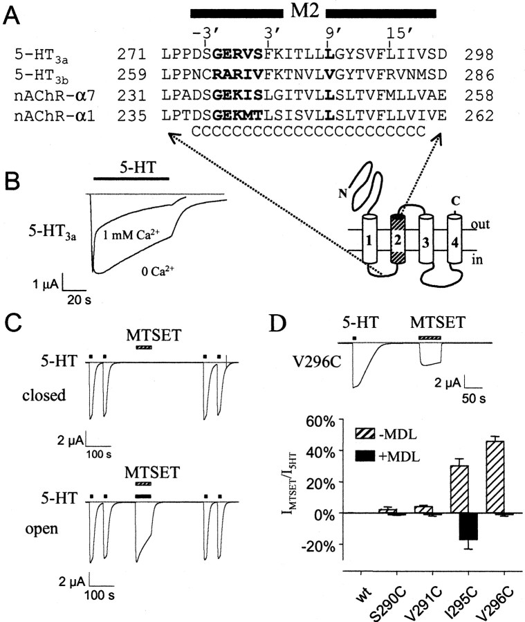
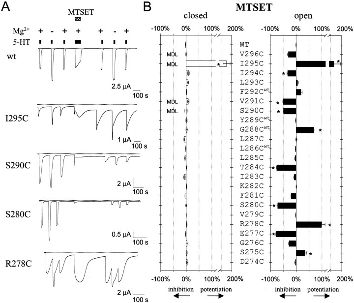
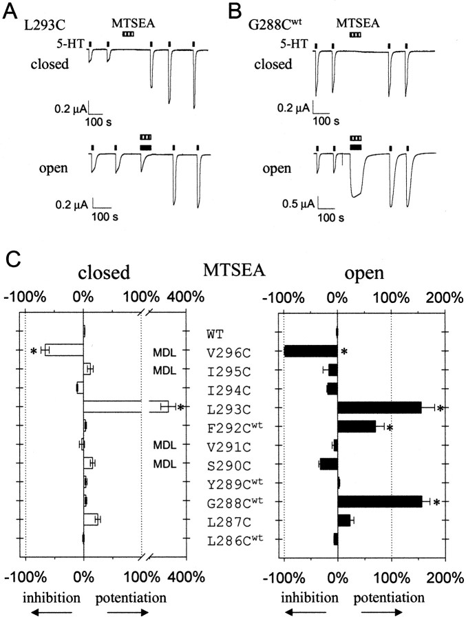
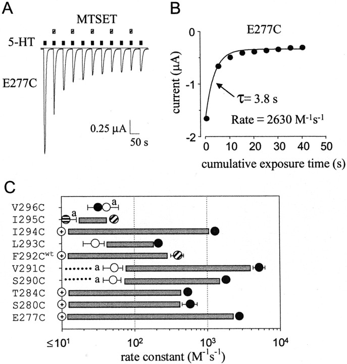
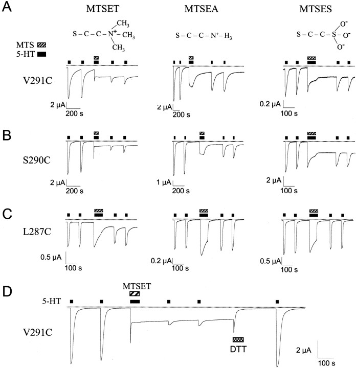
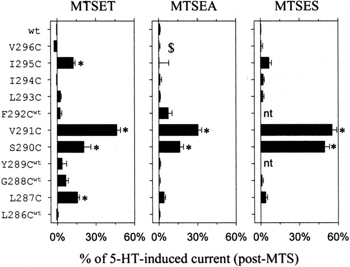
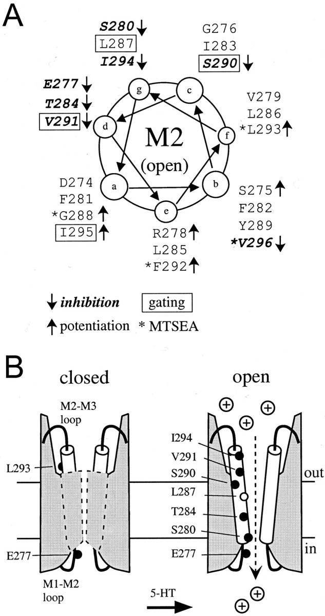
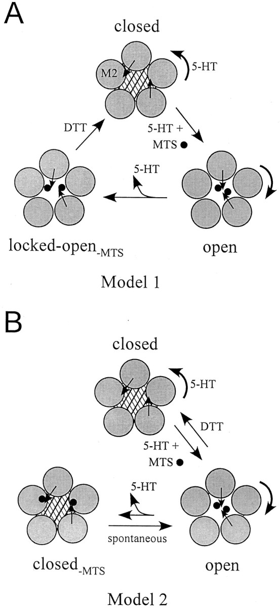
Serotonin-gated ion channels (5-HT3) are members of the ligand-gated channel family, which includes channels that are opened directly by the neurotransmitter acetylcholine, GABA, glycine, or glutamate. Although there is general agreement that the second transmembrane domain (M2) lines the pore, the position of the gate in the M2 is less certain. Here, we used substituted cysteine accessibility method (SCAM) to provide new evidence for a centrally located gate that moves during channel activation. In the closed state, three cysteine substitutions, located on the extracellular side of M2, were modified by methanethiosulfonate (MTS) reagents. In contrast, 13 cysteine substitutions were modified in the open state with MTS reagents. The pattern of inhibition (every three to four substitutions) was consistent with an alpha helical structure for the middle and cytoplasmic segments of the M2 transmembrane domain. Unexpectedly, open-state modification of two amino acids in the center of M2 with three different MTS reagents prevented channels from fully closing in the absence of neurotransmitter. Our results are consistent with a model in which the central region of the M2 transmembrane domain is inaccessible in the closed state and moves during channel activation.


Figures

Fig. 1.
Fig. 1.
Properties of wild-type and cysteine-substituted 5-HT3a channels expressed in Xenopusoocytes. A, Alignment of the M2 transmembrane domain sequences from mouse 5HT3a (M74425), mouse 5-HT3b (AF155045), mouse nAChRα7 (P49582), and mouse nAChRα1 (M17640) subunits. “C” indicates cysteine substitutions.Bold delimits two possible positions for the gate in nAChR, −2′G through 2′T (Akabas et al., 1994; Wilson and Karlin, 1998) and 9′L (Unwin, 1995). The proposed membrane topology is shown below.B, Removal of extracellular Ca2+slowed the rate of desensitization with 5-HT (10 μm).Dashed line indicates zero current level. All currents were recorded at −80 mV. C, Exposure of wild-type 5-HT3a channels to 1 mm MTSET in the closed or open (+10 μm 5-HT) state did not irreversibly change 5-HT-induced current. D, Continuous current recording from oocyte expressing V296C shows the direct activation with 1 mm MTSET. Bar graph shows the agonist activity (MTSET-induced current divided by the 5-HT-induced current) of 1 mm MTSET for S290C, V291C, I295C, and V296C (striped bar). The 5-HT3a antagonist MDL-72222 (1 μm; MDL) suppressed the agonist activity of MTSET (black bar) and MTSEA (data not shown). For I295C, MDL-72222 plus MTSET decreased the inward current, giving rise to a negative percentage of 5-HT-induced current.
Fig. 2.
Fig. 2.
Reactivity of cysteine-substituted 5-HT3a channels in open and closed states with MTSET.A, Continuous current recorded from oocyte injected with cRNA for wild-type, I295C, S290C, S280C, or R278C. Solid bar indicates 10 μm 5-HT, hatched bar indicates 1 mm MTSET, and + and − refers to extracellular solution with (+) or without (−) Mg2+. MTSET exposure irreversibly inhibited the 5-HT-induced current for S290C and S280C. The 5-HT-induced current for R278C increased in the +Mg2+ but not the −Mg2+ solution after MTSET treatment. I295C appeared to have slower deactivation kinetics after MTSET treatment (see Table 1). B, Summary of the percentage inhibition or potentiation after 1 min exposure to MTSET in either the closed (open bars) or open (solid bars) states (n = 4–17). L286Cwt, G288Cwt, Y289Cwt, and F292Cwt designate coexpression with wild-type 5-HT3a. MTSET was coapplied with 1 μmMDL-72222 to study closed-state inhibition for S290C, V291C, I295C, and V296C (MDL). The 5-HT-induced current was adjusted for desensitization for S290C (∼12%), V291C (∼16%), I294C (∼8%), and I295C (∼6%) that occurred after 1 min of 5-HT stimulation alone. *p < 0.05 indicates statistical difference from wild-type channels using one-way ANOVA.
Fig. 3.
Fig. 3.
Reactivity of cysteine-substituted 5-HT3a channels in open and closed states with MTSEA.A, B, Continuous current recorded from oocyte injected with the cRNA for L293C or G288Cwt. The 5-HT-induced current through L293C was potentiated with MTSEA (1 mm) in the closed and open states. G288Cwt was modified only in the open state.C, Summary of the percentage of inhibition or potentiation of current for mutants L286Cwt through V296C after 1 min exposure to MTSEA (1 mm) in the closed (open bars) or open (solid bars) states (n = 4–10). V296C and L293C were modified in both the closed and open states. The inhibition or potentiation for S290C, V291C, I294C, I295C, and V296C were adjusted as described in Figure 2. *p < 0.05 indicates statistical difference from wild-type channels using one-way ANOVA.
Fig. 4.
Fig. 4.
Rate constants for gated access to cysteine substitutions in the M2 transmembrane domain of 5-HT3a.A–C, Determination of the rate constant for modification with MTSET or MTSEA. A, Continuous current recorded from oocyte injected with the cRNA for E277C. Alternating pulses of 5-HT alone and 5-HT with MTSET (0.1 mm) were delivered. B, The amplitude of 5-HT-induced current was plotted as a function of cumulative exposure time to MTSET and fit with a single exponential having a time constant of 3.8 sec.C, Average rate constants for modification with MTSET (whitecircles, blackcircles) or MTSEA (circleswithhorizontal stripes, circles with diagonal stripes) in the closed and open states, respectively (n = 4–6). Circles with crosses indicate that closed-channel modification was not statistically different from wild type and was likely ≤10m/sec (see Fig. 2B). Note that closed rates are overestimates for those mutants in which MTSET opened the channel directly, denoted by a. Dashed line for S290C and V291C indicates there was no detectable effect of MTSET or MTSEA when coapplied with MDL-72222.
Fig. 5.
Fig. 5.
MTS modification in the open state produces agonist-independent currents for cysteine substitutions in the middle of the M2 transmembrane domain. Continuous current recorded from oocyte injected with the cRNA for V291C (A), S290C (B), or L287C (C). One minute exposure to MTS reagent (1 mm) in the presence of 5-HT (10 μm) produced an agonist-independent current for L287C, S290C, V291C, and I295C (data not shown) with MTSET and for S290C and V291C with MTSEA and MTSES. The structure of MTS moiety that would covalently attach to the cysteine sulfhydryl is shown above.D, Continuous current recorded from oocyte injected with cRNA for V291C. In the absence of 5-HT, 50 mm DTT decreased the agonist-independent current to the level before MTSET modification. Note that 5-HT (10 μm) fully activated the V291C channel after DTT treatment.
Fig. 6.
Fig. 6.
Summary of the change in agonist-independent current produced by MTSET, MTSEA, and MTSES. Bar graphs show the average amplitude of agonist-independent current after MTS treatment, expressed as a percentage of the 5-HT-induced current after MTS modification (see Materials and Methods) (n = 3–13). $ indicates that the pre-modified 5-HT-induced current was used to calculate the percentage change in agonist-independent current because MTSEA treatment eliminated >95% of the 5-HT-induced current. * indicates statistical difference from wild type using one-way ANOVA. nt, Not tested.
Fig. 7.
Fig. 7.
A, Helical wheel representation of the MTS-dependent changes in current. Note the majority of cysteine substitutions that are inhibited with MTS reagents (boldand italicized, ↓) fall along the same side of the helix, as do those that are potentiated (↑). Mutants that are locked open are boxed; mutants modified by MTSEA have anasterisk. B, Schematic model shows the extent of the gate in 5-HT3a, which is intracellular to L293. For clarity, only two of five subunits are shown with the M2 transmembrane domain. The M2 is modeled as a straight α helix in an open channel (the structure of M2 in the closed state is unknown). To explain the MTS accessibility of four cysteine substitutions located on the extracellular side of F292, the α helix is postulated to be water accessible on more than one face of the helix. The dashed area indicates that these cysteine substitutions (see Figs.2B, 3C) are accessible in the open but not closed state, suggesting that the gate extends from F292 to a more intracellular residue whose position can be determined by application of intracellular MTS. In the open state, the modification of cysteine substitutions leading to an inhibition of current are shown (black circles). L287C (9′) (white circle) was implicated previously in gating of nAChR. The narrowest region of the pore in nAChR is between E277 and S280 (Corringer et al., 2000).
Fig. 8.
Fig. 8.
Two working models that explain the agonist-independent current. In both models, 5-HT produces a rotation of M2 transmembrane domain, thereby exposing an ionized sulfhydryl on the cysteine (black arrow), such as at position V291 (only two cysteines are shown for clarity). MTS (black sphere) covalently attaches to the sulfhydryl in the open state (open). A, After removal of 5-HT, the channel fails to close completely in Model 1 because of steric hindrance (locked open-MTS). B, In Model 2, modified channel closes fully in the absence of 5-HT. This closed-MTS state is unstable, however, allowing the channel to spontaneously open. In both models, DTT can reduce the disulfide to restore the channel to its original unmodified closed state.

Similar articles

Cited by

References

    1. Akabas MH, Kaufmann C, Archdeacon P, Karlin A. Identification of acetylcholine receptor channel-lining residues in the entire M2 segment of the α subunit. Neuron. 1994;13:919–927. - PubMed
    1. Alhaider AA, Lei SZ, Wilcox GL. Spinal 5-HT3 receptor-mediated antinociception: possible release of GABA. J Neurosci. 1991;11:1881–1888. - PMC - PubMed
    1. Alkadhi KA, Salgado-Commissariat D, Hogan YH, Akpaudo SB. Induction and maintenance of ganglionic long-term potentiation require activation of 5-hydroxytryptamine (5-HT3) receptors. J Physiol (Lond) 1996;496:479–489. - PMC - PubMed
    1. Ballesteros JA, Shi L, Javitch JA. Structural mimicry in G protein-coupled receptors: implications of the high-resolution structure of rhodopsin for structure-function analysis of rhodopsin-like receptors. Mol Pharmacol. 2001;60:1–19. - PubMed
    1. Bertrand S, Devillers-Thiery A, Palma E, Buisson B, Edelstein SJ, Corringer P-J, Changeux J-P, Bertrand D. Paradoxical allosteric effects of competitive inhibitors on neuronal α7 nicotinic receptor mutants. Mol Neurosci. 1997;8:3591–3596. - PubMed

Publication types

MeSH terms

LinkOut - more resources