

Characterization of central inhibitory muscarinic autoreceptors by the use of muscarinic acetylcholine receptor knock-out mice
- PMID: 11880500
- PMCID: PMC6758851
- DOI: 10.1523/JNEUROSCI.22-05-01709.2002
Characterization of central inhibitory muscarinic autoreceptors by the use of muscarinic acetylcholine receptor knock-out mice
Abstract
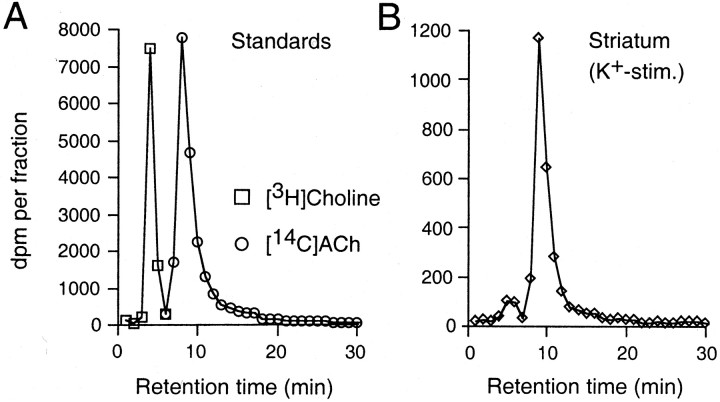
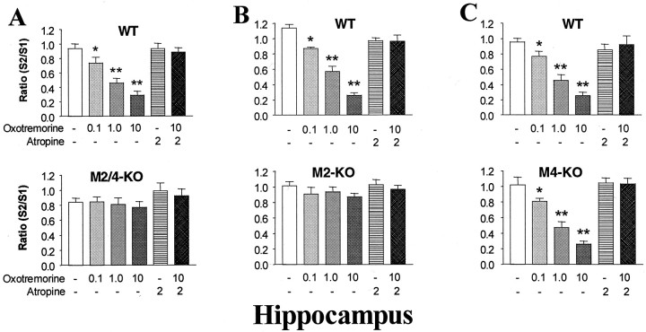
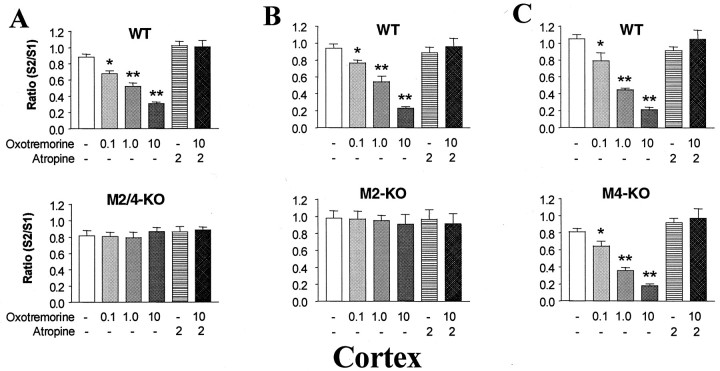
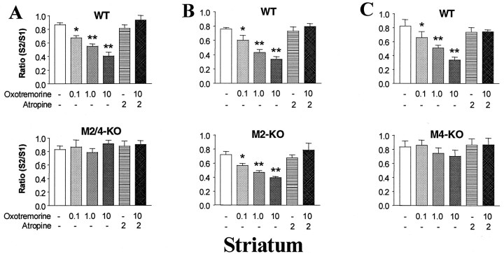
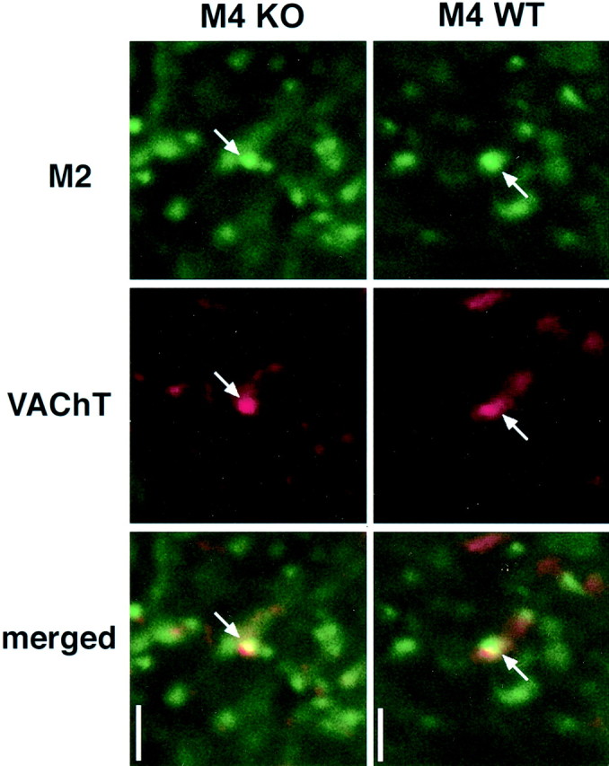
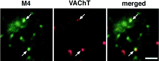
Forebrain muscarinic acetylcholine (ACh) receptors (mAChRs; M1-M5) are predicted to play important roles in many fundamental central functions, including higher cognitive processes and modulation of extrapyramidal motor activity. Synaptic ACh levels are known to be regulated by the activity of presynaptic muscarinic autoreceptors mediating inhibition of ACh release. Primarily because of the use of ligands with limited receptor subtype selectivity, classical pharmacological studies have led to conflicting results regarding the identity of the mAChR subtypes mediating this activity in different areas of the brain. To investigate the molecular identity of hippocampal, cortical, and striatal inhibitory muscarinic autoreceptors in a more direct manner, we used genetically altered mice lacking functional M2 and/or M4 mAChRs [knock-out (KO) mice]. After labeling of cellular ACh pools with [3H]choline, potassium-stimulated [3H]ACh release was measured in superfused brain slices, either in the absence or the presence of muscarinic drugs. The nonsubtype-selective muscarinic agonist, oxotremorine (0.1-10 microm), inhibited potassium-stimulated [3H]ACh release in hippocampal, cortical, and striatal slices prepared from wild-type mice by up to 80%. This activity was totally abolished in tissues prepared from M2-M4 receptor double KO mice. Strikingly, release studies with brain slices from M2 and M4 receptor single KO mice indicated that autoinhibition of ACh release is mediated primarily by the M2 receptor in hippocampus and cerebral cortex, but predominantly by the M4 receptor in the striatum. These results, together with additional receptor localization studies, support the novel concept that autoinhibition of ACh release involves different mAChRs in different regions of the brain.
Figures
Similar articles
-
Multiple muscarinic acetylcholine receptor subtypes modulate striatal dopamine release, as studied with M1-M5 muscarinic receptor knock-out mice.J Neurosci. 2002 Aug 1;22(15):6347-52. doi: 10.1523/JNEUROSCI.22-15-06347.2002. J Neurosci. 2002. PMID: 12151512 Free PMC article.
-
Modulation by adenosine of both muscarinic M1-facilitation and M2-inhibition of [3H]-acetylcholine release from the rat motor nerve terminals.Eur J Neurosci. 2002 Jun;15(11):1728-36. doi: 10.1046/j.1460-9568.2002.02020.x. Eur J Neurosci. 2002. PMID: 12081652
-
Striatal muscarinic receptors promote activity dependence of dopamine transmission via distinct receptor subtypes on cholinergic interneurons in ventral versus dorsal striatum.J Neurosci. 2010 Mar 3;30(9):3398-408. doi: 10.1523/JNEUROSCI.5620-09.2010. J Neurosci. 2010. PMID: 20203199 Free PMC article.
-
Use of M1-M5 muscarinic receptor knockout mice as novel tools to delineate the physiological roles of the muscarinic cholinergic system.Neurochem Res. 2003 Apr;28(3-4):437-42. doi: 10.1023/a:1022844517200. Neurochem Res. 2003. PMID: 12675128 Review.
-
Roles of the M4 acetylcholine receptor in the basal ganglia and the treatment of movement disorders.Mov Disord. 2019 Aug;34(8):1089-1099. doi: 10.1002/mds.27740. Epub 2019 Jun 18. Mov Disord. 2019. PMID: 31211471 Free PMC article. Review.
Cited by
-
Electron microscopic localization of M2-muscarinic receptors in cholinergic and noncholinergic neurons of the laterodorsal tegmental and pedunculopontine nuclei of the rat mesopontine tegmentum.J Comp Neurol. 2016 Oct 15;524(15):3084-103. doi: 10.1002/cne.24010. Epub 2016 Apr 21. J Comp Neurol. 2016. PMID: 27038330 Free PMC article.
-
Involvement of muscarinic receptors in psychomotor hyperactivity in dopamine-deficient mice.Mol Brain. 2022 Nov 29;15(1):96. doi: 10.1186/s13041-022-00984-x. Mol Brain. 2022. PMID: 36447257 Free PMC article.
-
AGAP1/AP-3-dependent endocytic recycling of M5 muscarinic receptors promotes dopamine release.EMBO J. 2010 Aug 18;29(16):2813-26. doi: 10.1038/emboj.2010.154. Epub 2010 Jul 27. EMBO J. 2010. PMID: 20664521 Free PMC article.
-
Characterization of muscarinic autoreceptors in the rabbit hippocampus and caudate nucleus.Neurochem Res. 2003 Apr;28(3-4):413-7. doi: 10.1023/a:1022836315383. Neurochem Res. 2003. PMID: 12675124
-
Xanomeline restores endogenous nicotinic acetylcholine receptor signaling in mouse prefrontal cortex.Neuropsychopharmacology. 2023 Mar;48(4):671-682. doi: 10.1038/s41386-023-01531-5. Epub 2023 Jan 12. Neuropsychopharmacology. 2023. PMID: 36635596 Free PMC article.
References
-
- Bernard V, Levey AI, Bloch B. Regulation of the subcellular distribution of m4 muscarinic acetylcholine receptors in striatal neurons in vivo by the cholinergic environment: evidence for regulation of cell surface receptors by endogenous and exogenous stimulation. J Neurosci. 1999;19:10237–10249. - PMC - PubMed
-
- Billard W, Binch H, III, Crosby G, McQuade RD. Identification of the primary muscarinic autoreceptor subtype in rat striatum as m2 through a correlation of in vivo microdialysis and in vitro receptor binding data. J Pharmacol Exp Ther. 1995;273:273–279. - PubMed
-
- Buckley NJ, Bonner TI, Buckley CM, Brann MR. Antagonist binding properties of five cloned muscarinic receptors expressed in CHO-K1 cells. Mol Pharmacol. 1989;35:469–476. - PubMed
Publication types
MeSH terms
Substances
Grants and funding
LinkOut - more resources
Full Text Sources
Molecular Biology Databases
Research Materials