. 2002 Mar 1;22(5):1738-51.
doi: 10.1523/JNEUROSCI.22-05-01738.2002.

Caspase-3-dependent proteolytic cleavage of protein kinase Cdelta is essential for oxidative stress-mediated dopaminergic cell death after exposure to methylcyclopentadienyl manganese tricarbonyl

Affiliations

Caspase-3-dependent proteolytic cleavage of protein kinase Cdelta is essential for oxidative stress-mediated dopaminergic cell death after exposure to methylcyclopentadienyl manganese tricarbonyl

Vellareddy Anantharam et al. J Neurosci. .

Abstract

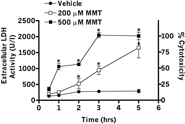
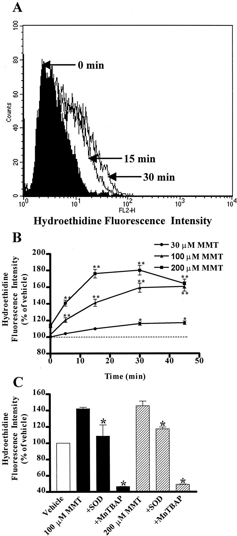
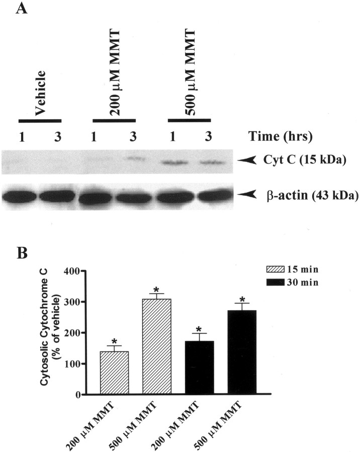
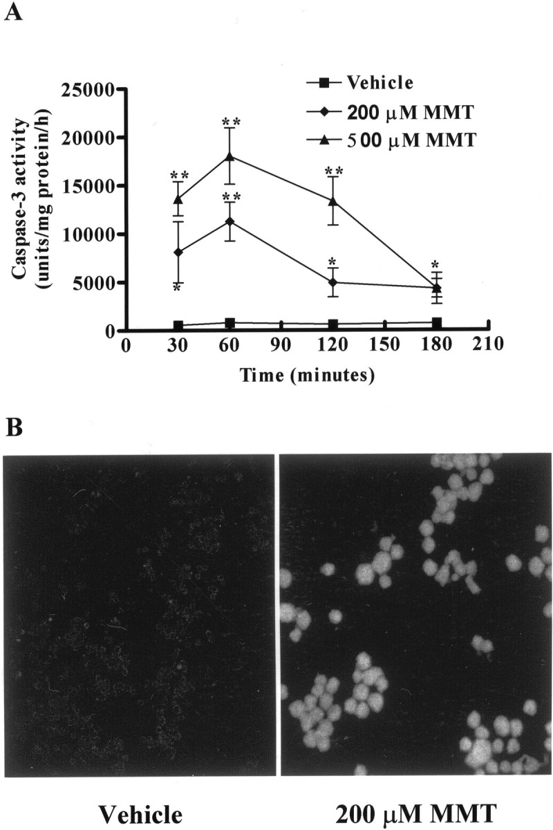
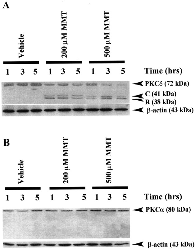
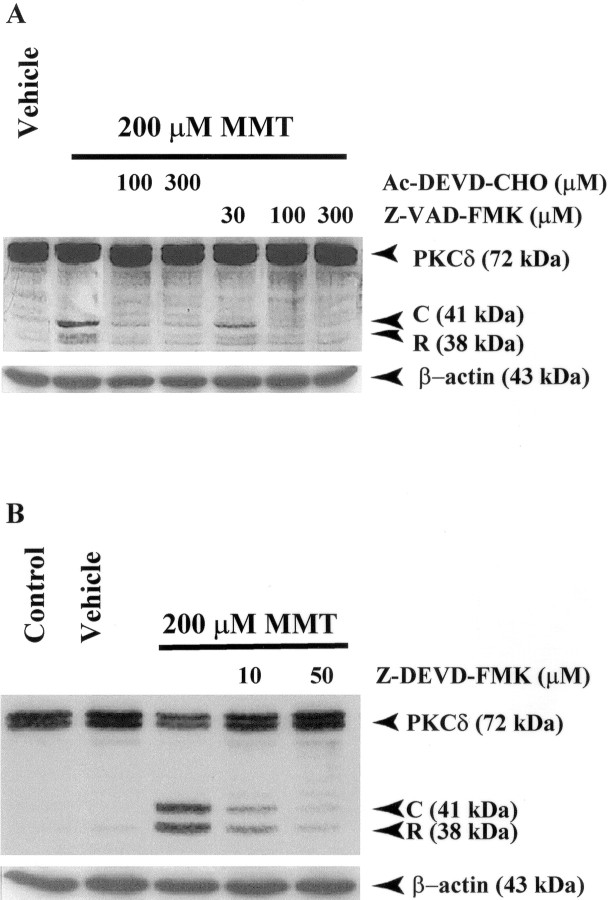
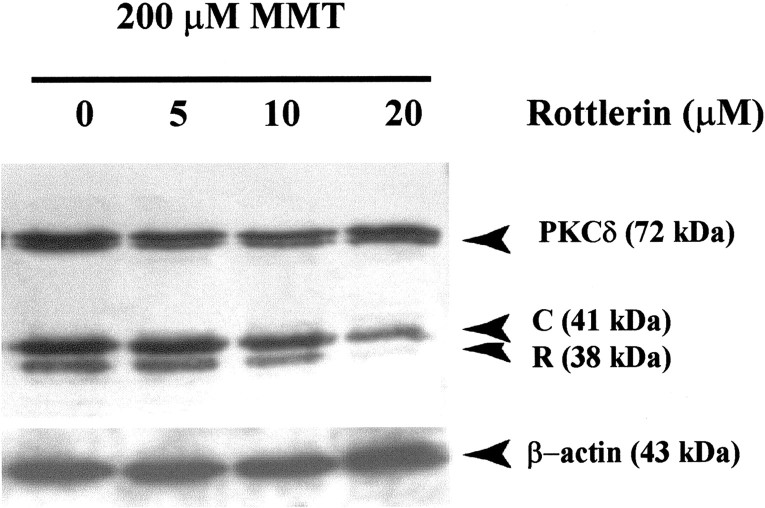
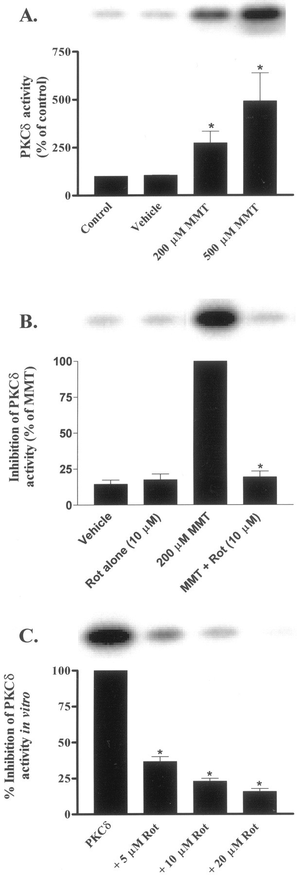
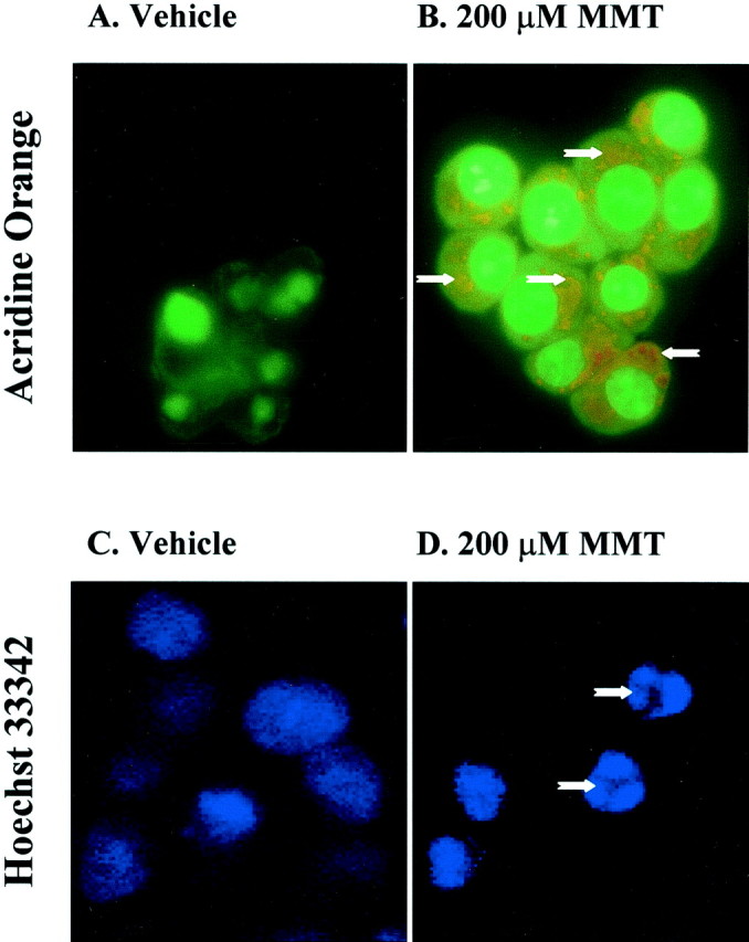
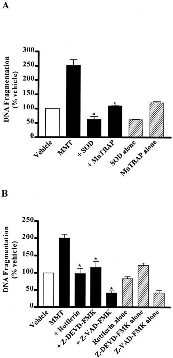
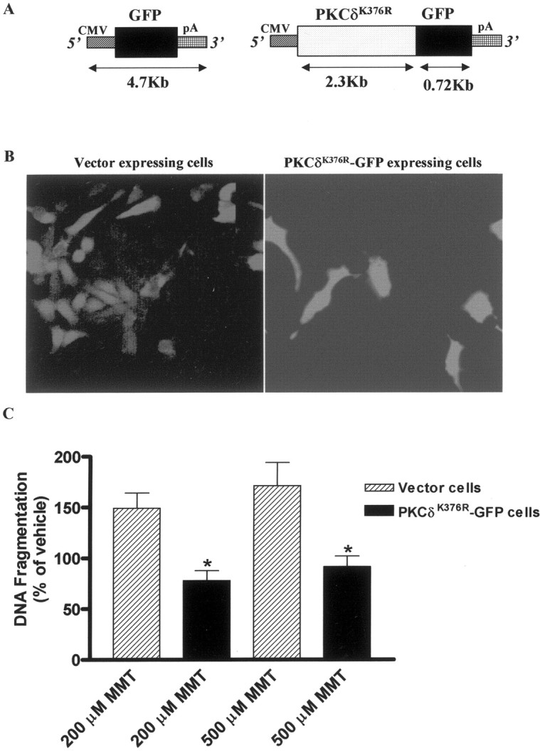
In the present study, we characterized oxidative stress-dependent cellular events in dopaminergic cells after exposure to an organic form of manganese compound, methylcyclopentadienyl manganese tricarbonyl (MMT). In pheochromocytoma cells, MMT exposure resulted in rapid increase in generation of reactive oxygen species (ROS) within 5--15 min, followed by release of mitochondrial cytochrome C into cytoplasm and subsequent activation of cysteine proteases, caspase-9 (twofold to threefold) and caspase-3 (15- to 25-fold), but not caspase-8, in a time- and dose-dependent manner. Interestingly, we also found that MMT exposure induces a time- and dose-dependent proteolytic cleavage of native protein kinase Cdelta (PKCdelta, 72-74 kDa) to yield 41 kDa catalytically active and 38 kDa regulatory fragments. Pretreatment with caspase inhibitors (Z-DEVD-FMK or Z-VAD-FMK) blocked MMT-induced proteolytic cleavage of PKCdelta, indicating that cleavage is mediated by caspase-3. Furthermore, inhibition of PKCdelta activity with a specific inhibitor, rottlerin, significantly inhibited caspase-3 activation in a dose-dependent manner along with a reduction in PKCdelta cleavage products, indicating a possible positive feedback activation of caspase-3 activity by PKCdelta. The presence of such a positive feedback loop was also confirmed by delivering the catalytically active PKCdelta fragment. Attenuation of ROS generation, caspase-3 activation, and PKCdelta activity before MMT treatment almost completely suppressed DNA fragmentation. Additionally, overexpression of catalytically inactive PKCdelta(K376R) (dominant-negative mutant) prevented MMT-induced apoptosis in immortalized mesencephalic dopaminergic cells. For the first time, these data demonstrate that caspase-3-dependent proteolytic activation of PKCdelta plays a key role in oxidative stress-mediated apoptosis in dopaminergic cells after exposure to an environmental neurotoxic agent.


Figures

Fig. 1.
Fig. 1.
MMT exposure induces cell death. PC12 cells were exposed to 200 and 500 μm of MMT for 0.5–5 hr at 37°C. After the exposure, cell-free extracellular supernatants were collected, and LDH activity was measured by spectrophotometer. Values represent mean ± SEM for three to five separate experiments in triplicate. Significance was determined by ANOVA followed by Dunnett's post-test between the vehicle-treated group and each treatment group (*p < 0.05).
Fig. 2.
Fig. 2.
MMT treatment generates ROS in PC12 cells. PC12 cells were suspended in HBSS supplemented with 2 mmCa2+ at a density of 0.5–0.75 × 10⁶ cells/ml. A concentration of 10 μmhydroethidine was added to the cells and incubated for 15 min at 37°C in the dark. A, Time-dependent change in hydroethidine fluorescent intensity in PC12 cells treated with MMT. A concentration of 200 μm MMT was added, and fluorescent intensity was measured at 0, 15, and 30 min by flow cytometry as described in Materials and Methods. The data are a representative flow cytometric histogram of MMT-treated PC12 cells exhibiting a time-dependent increase in red fluorescence. B, Dose- and time-dependent increase in ROS production. Various doses of MMT were added, and fluorescent intensity was measured at 0, 5, 15, and 30 min. Data represent the mean ± SEM of two to five separate experiments in triplicate. Asterisks (*p < 0.5 and **p < 0.01) indicate significant differences compared with the time-matched vehicle-treated cells. C, Effect of SOD and MnTBAP on ROS production. Cells were pretreated with ROS inhibitors, SOD (100 U/ml) and MnTBAP (10 μm), and then exposed to 100 or 200 μm MMT for 15 min. The value of each treatment group is the mean ± SEM from two to three separate experiments performed in triplicate. Asterisks (*p < 0.05) indicate significant differences compared with MMT-treated cells.
Fig. 3.
Fig. 3.
Dose- and time-dependent accumulation of cytosolic cytochrome C in MMT-treated PC12 cells. A, Western blot.B, Cytochrome C ELISA assay. A, Subconfluent cultures of undifferentiated PC12 cells were harvested at 1 and 3 hr after treatment with 200 or 500 μm MMT. The cytosolic fractions were obtained as described in Materials and Methods. Cytosolic fractions were separated by 12% SDS-PAGE, transferred to a nitrocellulose membrane, and cytochrome C (Cyt C) was detected using polyclonal antibody raised against cytochrome C. For β-actin measurements, the membrane used for cytochrome C was reprobed with β-actin antibody to confirm equal protein loading in each lane. The immunoblots were visualized using ECL detection agents from Amersham. B, Subconfluent cultures of undifferentiated PC12 cells were harvested at 15 and 30 min after treatment with 200 or 500 μm MMT. The cytosolic fractions were obtained as described in Materials and Methods. The value of each treatment group is the mean ± SEM from two separate experiments in triplicate. Asterisks (*p < 0.05) indicate significant differences compared with vehicle-treated cells.
Fig. 4.
Fig. 4.
MMT treatment increases caspase-3 activity.A, Caspase-3 enzymatic activity. B, In situ caspase-3 activity. A, Subconfluent cultures of undifferentiated PC12 cells were harvested at 30 min, 1, 2, and 3 hr after MMT treatment. Caspase-3 activity was assayed using specific fluorogenic substrate, Ac-DEVD-AMC (50 μm), as described in Materials and Methods. The data represent mean ± SEM of nine individual measurements from three separate experiments. Asterisks (**p < 0.01; *p < 0.05) indicate significant differences compared with temporally matched vehicle (DMSO)-treated cells. B, PC12 cells were grown on laminin-coated slides for 2–3 d and then exposed to 0.5% DMS0 (vehicle) and 200 μm MMT for 1 hr in the dark. After exposure, cells were treated with 10 μm FITC-VAD-FMK (Promega caspACE, in situ marker for caspase-3 activity) and processed as described in Materials and Methods. Confocal images were obtained using a Leica TCS-NT microscope.
Fig. 5.
Fig. 5.
Proteolytic cleavage of PKCδ but not of PKCα in MMT-treated PC12 cells. A, PKCδ; B,PKCα. Subconfluent undifferentiated PC12 cells were harvested at 1, 3, and 5 hr after treatment of 200 or 500 μm MMT. Cytosolic fractions were obtained as described in Materials and Methods, and were separated by 10% SDS-PAGE, transferred to nitrocellulose membrane, and PKCα and PKCδ were detected using antibodies directed against their catalytic subunits. To confirm equal protein loading in each lane, the membranes were reprobed with β-actin antibody. The immunoblots were visualized using ECL detection agents from Amersham. C, PKCδ catalytic subunit;R, PKCδ regulatory subunit.
Fig. 6.
Fig. 6.
Caspase-3 mediates the proteolytic cleavage of PKCδ in MMT-treated PC12 cells. A, Effect of Ac-DEVD-CHO and Z-VAD-FMK on PKCδ cleavage. B, Effect of Z-DEVD-FMK on PKCδ cleavage. Subconfluent undifferentiated PC12 cells were treated with 200 μm MMT, with or without the inclusion of caspase inhibitors Ac-DEVD-CHO, Z-VAD-FMK, or Z-DEVD-FMK. Inhibitors were added 30 min before the addition of MMT. Cells were harvested 3 hr after the addition of MMT. The cytosolic fractions were obtained as described in Materials and Methods, and were analyzed by 10% SDS-PAGE and Western blot. To confirm equal protein loading in each lane, the membranes were reprobed with β-actin antibody.C, PKCδ catalytic subunit; R, PKCδ regulatory subunit.
Fig. 7.
Fig. 7.
Suppression of caspase-3 activity by rottlerin in MMT-treated PC12 cells. A, Pre-treatment;B, post-treatment. Subconfluent undifferentiated PC12 cells were treated with 200 μm MMT with or without the inclusion of rottlerin (Rot; 5–20 μm) for 1 hr. Rottlerin was added 30 min before or 30 min after the addition of MMT. Caspase-3 activity was assayed using Ac-DEVD-AMC (50 μm) as substrate, as described in Materials and Methods. The data represent an average of four to nine individual measurements from two or three separate experiments ± SEM. Asterisk (*p < 0.05) indicates significant difference compared with cells exposed to 200 μm MMT.
Fig. 8.
Fig. 8.
Rottlerin pretreatment blocks proteolytic cleavage of PKCδ in MMT-treated PC12 cells. Subconfluent undifferentiated PC12 cells were treated with 200 μm MMT with or without the inclusion of rottlerin (5–20 μm). Rottlerin was added 90 min before the addition of MMT. Cytosolic fractions were obtained as described in Materials and Methods and were separated by 10% SDS-PAGE, transferred to nitrocellulose membrane, and PKCδ was detected using an antibody directed against its catalytic subunit. The immunoblots were visualized using ECL detection agents from Amersham. To confirm equal protein loading in each lane, the membranes were reprobed with β-actin antibody. C, PKCδ catalytic subunit;R, PKCδ regulatory subunit.
Fig. 9.
Fig. 9.
Rottlerin inhibits PKCδ kinase activity in intact cells and in in vitro. A, Dose-dependent increase in PKCδ activity. B, Rottlerin suppresses MMT-induced increase in PKCδ kinase activity in intact cells. C, Rottlerin inhibits PKCδ kinase activityin vitro. Subconfluent undifferentiated PC12 cells were treated with 200 μm MMT for 1 hr at 37°C with or without the inclusion of rottlerin (Rot; 5–20 μm). Rottlerin was added 30 min before the addition of MMT. For in vitro inhibition of PKCδ activity, rottlerin (5–20 μm) was added to the immunoprecipitated samples from MMT-treated cells and incubated for 30 min before the addition of substrate (histone H1) and [γ-32P]ATP. The immunoprecipitation kinase assay was performed as described in Materials and Methods. The bands were quantified by a PhosphoImager after scanning the dried gel and expressed as a percentage of control (untreated cells) (A), percentage of MMT treatment (B), or percentage of PKCδ kinase activity (C). The data represent an average of three individual measurements from two separate experiments ± SEM. Asterisks (*p < 0.05) indicate significant differences compared with control, MMT-treated cells, or PKCδ kinase activity.
Fig. 10.
Fig. 10.
MMT treatment increases apoptosis in situ. A, C, Vehicle-treated cells; B, D, 200 μm MMT-treated cells. PC12 cells were grown on laminin-coated slides for 2–3 d and then exposed to 200 μm MMT for 1 hr. A, B, For acridine orange staining, cells were treated with acridine orange (10 μm) for 15 min in the dark at RT after exposure to MMT.Arrows indicate enhanced red fluorescence and reduced green fluorescence in MMT-treated cells, which are undergoing apoptosis, whereas little or no enhanced redfluorescence was seen in vehicle-treated cells. C, D,For Hoechst 33342 staining, cells were stained with Hoechst 33342 (10 μg/ml) for 3 min in dark after exposure to MMT. Arrowsindicate apoptotic cells containing condensed chromatin.
Fig. 11.
Fig. 11.
Suppression of MMT-induced apoptosis in PC12 cells. A, ROS inhibitors, SOD and MnTBAP.B, Caspase-3 inhibitors, Z-VAD-FMK and Z-DEVD-FMK and PKCδ inhibitor, rottlerin. Subconfluent cultures of undifferentiated PC12 cells were treated with MMT (200 μm) with or without the inclusion of the following inhibitors: ROS inhibitors SOD (100 U/ml) or MnTBAP (10 μm); caspase inhibitors Z-VAD-FMK (100 μm) or Z-DEVD-FMK (50 μm); and PKCδ inhibitor rottlerin (10 μm). Inhibitors were added 30 min before addition of MMT. Cells were harvested 1 hr after MMT treatment. Apoptosis was assayed using ELISA assay as described in Materials and Methods. The data are expressed as percentage of apoptosis observed in vehicle-treated cells. The data represent the mean ± SEM of six individual measurements from three separate experiments. Asterisks (*p < 0.01) indicate significant differences when compared with cells exposed to 200 μm MMT.
Fig. 12.
Fig. 12.
Overexpression of catalytically inactive PKCδ protein blocks MMT-induced apoptosis in immortalized dopaminergic neuronal cell line (1RB3AN27).A, Plasmid description, pEGFP-NI construct codes for the green fluorescent protein (GFP) mRNA transcribed under the 5′ human cytomegalovirus (CMV) immediate early promoter, and the mRNA is stabilized with the 3′ SV40 mRNA polyadenylation signal (pA) and was used as vector control. PKCδK376R-GFP construct codes for the kinase inactive PKCδ-GFP fusion transcript. B, Stable expression of GFP and PKCδK376R-GFP fusion protein in 1RB3AN27 cells. The cells were viewed under a fluorescence microscope, and images were obtained with a SPOT digital camera. C, Subconfluent cultures of undifferentiated 1RB3AN27 cells stably expressing vector or PKCδK376R-GFP fusion protein were treated with MMT (200 and 500 μm) for 3 hr. Apoptosis was assayed using ELISA assay as described in Materials and Methods. The data are expressed as percentage of apoptosis observed in vehicle-treated cells. The data represent a mean ± SEM of four to six individual measurements from two separate experiments. Asterisks (*p < 0.01) indicate significant differences when compared with MMT-treated cells.
Fig. 13.
Fig. 13.
A model describing the sequence of cell death signaling events in MMT-induced apoptosis. 1, Increased ROS production can be blocked by pretreatment with antioxidants, superoxide dismutase and MnTBAP; 2, cytochrome C is released into the cytosol from the mitochondria; 3,cytosolic cytochrome C activates caspase-9; 4, caspase-9 activates caspase-3; 5, caspase-3 mediates proteolytic cleavage of PKCδ, which can be blocked by pretreatment with the caspase inhibitors Ac-DEVD-CHO, Z-VAD-FMK, and Z-DEVD-FMK;6, pretreatment with rottlerin, a PKCδ inhibitor, reduces caspase-3 activity indicating a possible feedback activation;7, both caspase-3 and PKCδ inhibitors block MMT-induced DNA fragmentation; and 8, dopaminergic cells stably overexpressing catalytically inactive PKCδ [dominant-negative mutant (DNM) PKCδK376RGFP] completely blocked MMT-induced DNA fragmentation. In conclusion, our data suggest that caspase-3-dependent proteolytic activation of PKCδ plays a key role in MMT-induced dopaminergic cell death.

Similar articles

Cited by

References

    1. Ajiro K. Histone H2B phosphorylation in mammalian apoptotic cells An association with DNA fragmentation. J Biol Chem. 2000;275:439–443. - PubMed
    1. Aschner M. Manganese brain transport and emerging research needs. Environ Health Perspect. 2000;108 [Suppl 3]:429–432. - PMC - PubMed
    1. Autissier N, Dumas P, Brosseau J, Loireau A. Effects of methylcyclopentadienyl manganese tricarbonyl (MMT) of rat liver mitochondria. I. Effects, in vitro, on the oxidative phosphorylation. Toxicology. 1977;7:115–122. - PubMed
    1. Barbeau A. Manganese and extrapyramidal disorders (a critical review and tribute to Dr. George C Cotzias). Neurotoxicology. 1984;5:13–35. - PubMed
    1. Basu A, Woolard MD, Johnson CL. Involvement of protein kinase C-delta in DNA damage-induced apoptosis. Cell Death Differ. 2001;8:899–908. - PubMed

Publication types

MeSH terms

Substances