

Retrograde repression of growth-associated protein-43 mRNA expression in rat cortical neurons
- PMID: 11880510
- PMCID: PMC6758898
- DOI: 10.1523/JNEUROSCI.22-05-01816.2002
Retrograde repression of growth-associated protein-43 mRNA expression in rat cortical neurons
Abstract
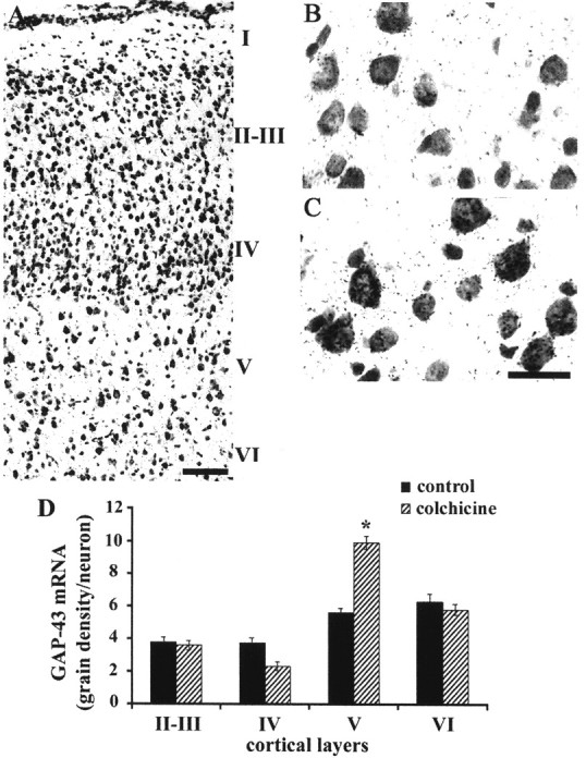
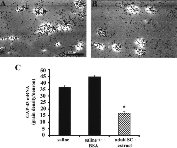
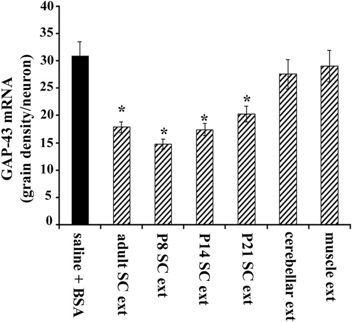
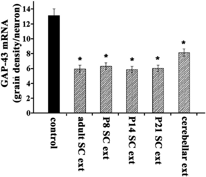
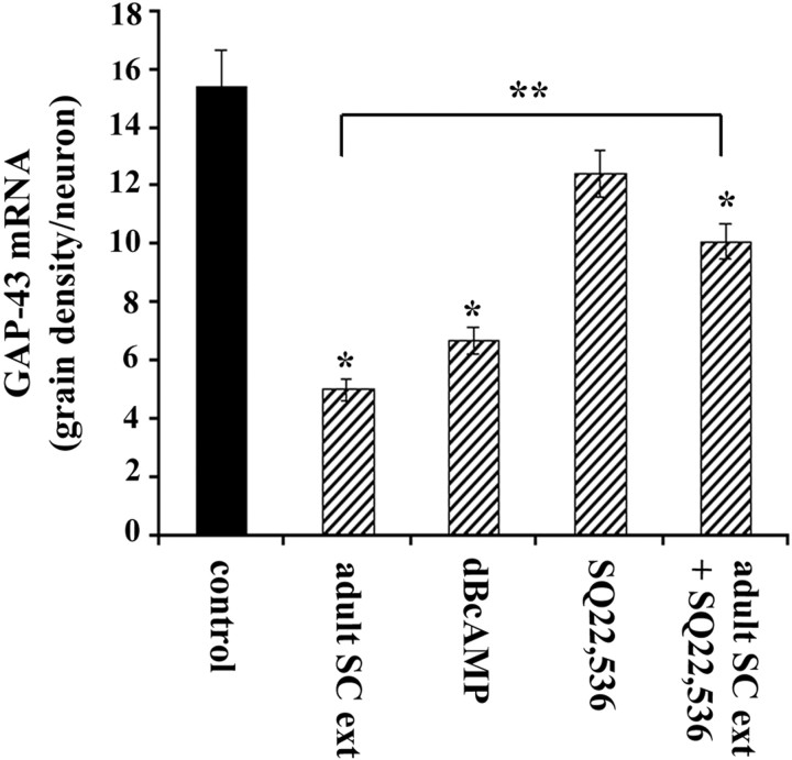
Corticospinal neurons support rapid growth of axons toward spinal cord targets in the perinatal period. Initial axon growth is accompanied by elevated expression of growth-associated protein-43 (GAP-43), which then declines in postnatal development. To investigate whether expression of GAP-43 mRNA is regulated by retrograde signals, we injected colchicine into the corticospinal tract to block retrograde axonal transport during a time when GAP-43 is normally declining in corticospinal neurons. Colchicine caused a prolongation of high GAP-43 mRNA expression in neurons located in layer V (but not other layers) of sensorimotor cortex. We next used osmotic minipumps to infuse soluble adult spinal cord extract into the sensorimotor cortex. This resulted in a premature downregulation of GAP-43 mRNA in identified corticospinal neurons. GAP-43 repressive activity was found in extracts of the spinal cord tissue as young as postnatal day 8. The effect of spinal cord extract in vivo was not mimicked by adult cerebellar or muscle extracts. Cultures of postnatal cortical neurons also underwent downregulation of GAP-43 mRNA when treated with spinal cord extract. Activation of cAMP signaling also repressed GAP-43 mRNA in cortical cultures, and the repressive effect of spinal cord extract was diminished by an adenyl cyclase inhibitor. Thus, GAP-43 mRNA may be downregulated late in development by a target-derived retrograde repressive factor, and this effect may be mediated by cAMP second messenger signaling.
Figures
Similar articles
-
Developmental down-regulation of GAP-43 expression and timing of target contact in rat corticospinal neurons.Exp Neurol. 2002 Aug;176(2):390-401. doi: 10.1006/exnr.2002.7964. Exp Neurol. 2002. PMID: 12359181
-
Cyclic AMP prevents an increase in GAP-43 but promotes neurite growth in cultured adult rat dorsal root ganglion neurons.Exp Neurol. 2000 Nov;166(1):153-65. doi: 10.1006/exnr.2000.7485. Exp Neurol. 2000. PMID: 11031091
-
Increased expression of the growth-associated protein 43 gene in the sensorimotor cortex of the macaque monkey after lesioning the lateral corticospinal tract.J Comp Neurol. 2009 Oct 20;516(6):493-506. doi: 10.1002/cne.22121. J Comp Neurol. 2009. PMID: 19672995
-
2003 Keio Medical Science Prize commemorative lecture. Neural mechanisms of cognitive memory.Keio J Med. 2004 Jun;53(2):59-68. doi: 10.2302/kjm.53.59. Keio J Med. 2004. PMID: 15264368 Review. No abstract available.
-
Viral assembly using heterologous expression systems and cell extracts.Adv Protein Chem. 2003;64:1-36. doi: 10.1016/s0065-3233(03)01001-5. Adv Protein Chem. 2003. PMID: 13677044 Review. No abstract available.
Cited by
-
Cell-autonomous mechanisms and myelin-associated factors contribute to the development of Purkinje axon intracortical plexus in the rat cerebellum.J Neurosci. 2003 Jun 1;23(11):4613-24. doi: 10.1523/JNEUROSCI.23-11-04613.2003. J Neurosci. 2003. PMID: 12805301 Free PMC article.
-
Examination of the combined effects of chondroitinase ABC, growth factors and locomotor training following compressive spinal cord injury on neuroanatomical plasticity and kinematics.PLoS One. 2014 Oct 28;9(10):e111072. doi: 10.1371/journal.pone.0111072. eCollection 2014. PLoS One. 2014. PMID: 25350665 Free PMC article.
-
Flipping the transcriptional switch from myelin inhibition to axon growth in the CNS.Front Mol Neurosci. 2015 Jul 17;8:34. doi: 10.3389/fnmol.2015.00034. eCollection 2015. Front Mol Neurosci. 2015. PMID: 26236189 Free PMC article.
-
cJun promotes CNS axon growth.Mol Cell Neurosci. 2014 Mar;59:97-105. doi: 10.1016/j.mcn.2014.02.002. Epub 2014 Feb 9. Mol Cell Neurosci. 2014. PMID: 24521823 Free PMC article.
References
-
- Andersen PL, Webber CA, Kimura KA, Schreyer DJ. Cyclic AMP prevents an increase in GAP-43 but promotes neurite growth in cultured adult rat dorsal root ganglion neurons. Exp Neurol. 2000a;166:153–165. - PubMed
-
- Andersen PL, Webber CA, Whittemore SR, Schreyer DJ. Divergent regulation of GAP-43 expression and CNS neurite outgrowth by cyclic AMP. J Neurosci Res. 2000b;61:626–635. - PubMed
-
- Basi GS, Jacobson RD, Virag I, Schilling J, Skene JH. Primary structure and transcriptional regulation of GAP-43, a protein associated with nerve growth. Cell. 1987;49:785–791. - PubMed
Publication types
MeSH terms
Substances
LinkOut - more resources
Full Text Sources
Miscellaneous