

. 2002 Apr 30;99 Suppl 2(Suppl 2):6451-5.
doi: 10.1073/pnas.221458298.
Epub 2002 Mar 5.
Emulating biology: building nanostructures from the bottom up
Affiliations
- PMID: 11880609
- PMCID: PMC128548
- DOI: 10.1073/pnas.221458298
Item in Clipboard
Emulating biology: building nanostructures from the bottom up
Proc Natl Acad Sci U S A.
.
Abstract
The biological approach to nanotechnology has produced self-assembled objects, arrays and devices; likewise, it has achieved the recognition of inorganic systems and the control of their growth. Can these approaches now be integrated to produce useful systems?
Figures
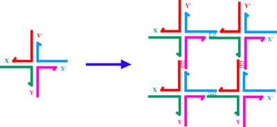
Formation of a 2D lattice from a junction with sticky ends. X and Y are sticky ends and X′ and Y′ are their complements. Four of the monomers on the left are complexed to yield the structure on the right. DNA ligase can close the gaps left in the complex, which can be extended by the addition of more monomers.
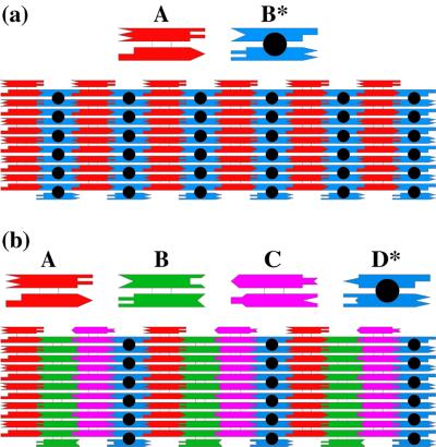
Arrays assembled from DX molecules. (a) A two-component array. Two DX molecules (A and B*) are illustrated schematically (a Top). The two helices are drawn as rectangles, and the complementary sticky ends are represented by geometrical shapes. A is a conventional DX molecule, but B* contains a DNA hairpin protruding from the plane. Below these molecules is an array that shows the two components fitting together to tile a plane. (b) A four-component array. The same conventions apply as in a. This array uses four tiles, A, B, C, and D*, where A, B, and C are conventional DX molecules and D* contains a hairpin. The stripes are separated by twice the distance seen in a.
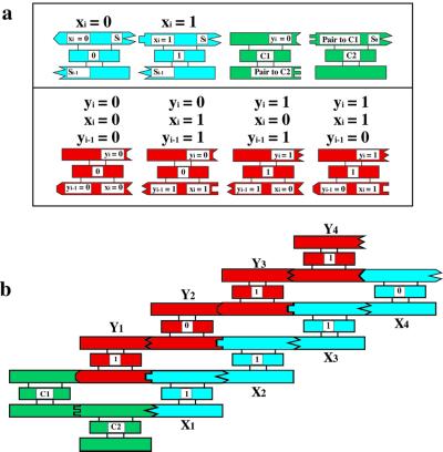
A cumulative XOR calculation. The XOR operation takes two Boolean inputs and produces a 0 if they are the same and a 1 if they are different. Shown in a are blue input tiles, Xi which represent 0 or 1, according to the presence of a particular restriction enzyme site. These have been assembled in a particular order in b. The red tiles in a contain the four Boolean possibilities as sticky ends on their lower helices. The input is connected to the output through the green C1 and C2 tiles. At the end of the self-assembly, one strand that runs through the entire system is ligated together, thereby connecting the input to the output. It is read by partial restriction, followed by a denaturing gel, much like a sequencing reaction.
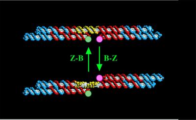
A DNA nanomechanical device based on the B–Z transition. The device consists of two DX molecules connected by a helix (yellow section) that can undergo the B-Z transition. When this occurs, the bottom domain of the right DX molecule swings from the bottom to the top through a rotary motion.
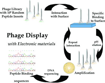
Peptide selection for electronic materials. The 1.9 × 10⁹ random peptide sequences are exposed to the different crystal substrates; nonspecific peptide interactions are removed with extensive washes. The phage that bind are eluted by lowering the pH and disrupting the surface interaction. The eluted phage are amplified by infecting the E. coli ER2537 host, producing enriched populations of phage, displaying peptides that interact with the specific crystal substrate. The amplified phage are isolated, titered, and reexposed to a freshly prepared substrate surface, thereby enriching the phage population with substrate-specific binding phage. This procedure is repeated three to five times to select the phage with the tightest and most specific binding. The DNA of phage that show specificity is sequenced to determine the peptide-binding sequence.
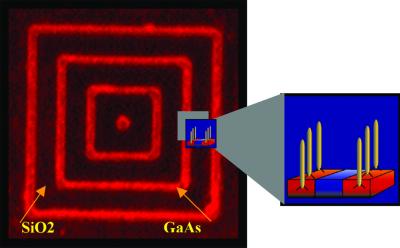
Peptide selectivity for patterned GaAs. Fluorescently labeled phage displaying a peptide specifically selected to bind to GaAs bind only to the patterned GaAs nested square pattern on a wafer. The red lines (1 μm across) correspond to GaAs and the black spaces (4 μm across) are SiO2. This peptide-specific binding could also be used to deliver nanocrystals to specific locations.
References
-
- Seeman N C. Trends Biotechnol. 1999;17:437–443. - PubMed
-
- Seeman N C. J Theor Biol. 1982;99:237–247. - PubMed
-
- Qiu H, Dewan J C, Seeman N C. J Mol Biol. 1997;267:881–898. - PubMed
-
- Robinson B H, Seeman N C. Protein Eng. 1987;1:295–300. - PubMed
-
- Petrillo M L, Newton C J, Cunningham R P, Ma R-I, Kallenbach N R, Seeman N C. Biopolymers. 1988;27:1337–1352. - PubMed
Publication types
MeSH terms
Substances
Grants and funding
LinkOut - more resources
Full Text Sources
Other Literature Sources
