. 2002 Mar 5;99(5):2989-94.
doi: 10.1073/pnas.052714099.

Different contributions of thymopoiesis and homeostasis-driven proliferation to the reconstitution of naive and memory T cell compartments

Affiliations

Different contributions of thymopoiesis and homeostasis-driven proliferation to the reconstitution of naive and memory T cell compartments

Qing Ge et al. Proc Natl Acad Sci U S A. .

Abstract

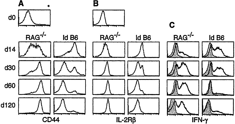
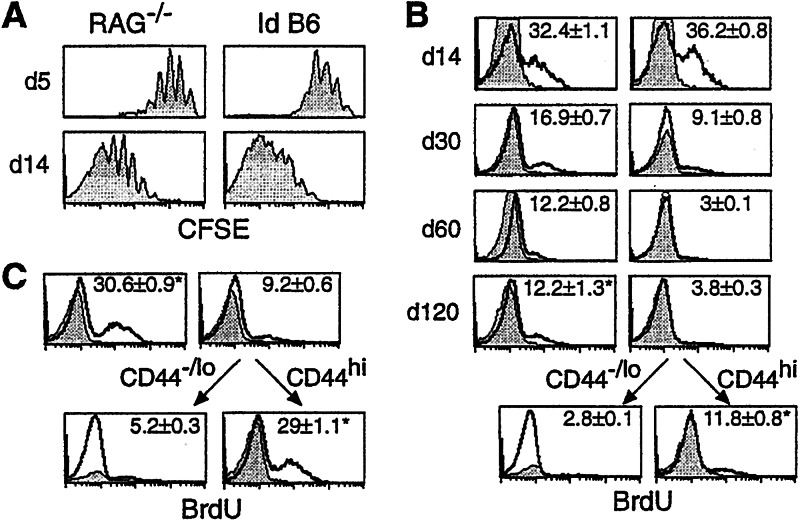
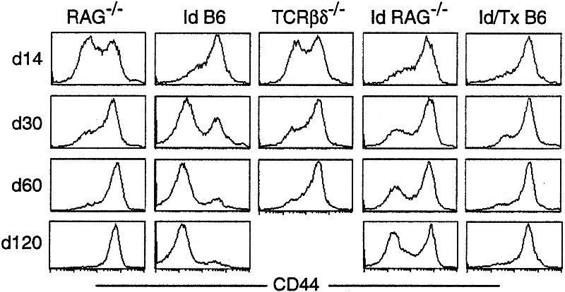
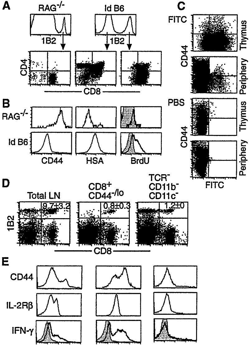
Following transfer into lymphopenic hosts, naive CD8 T cells proliferate and acquire memory phenotype. Although the acquired phenotype is stable in recombination activating gene-1-deficient (RAG-/-) recipients, in sublethally irradiated mice naive CD8 T cells of donor origin gradually accumulate. The naive cells have been attributed to phenotypic reversion of homeostatic memory cells, implying instability of memory phenotype and restoration of the naive T cell compartment by homeostasis-driven proliferation. We show here that (i) the accumulation of naive CD8 T cells of donor origin only occurs in recipients that have been irradiated and have an intact thymus; (ii) the apparent reversion of memory to naive cells actually results from de novo T cell development of hematopoietic stem cells, present in the donor spleen or lymph node cell populations, in the thymus of irradiated recipients; and (iii) the number of homeostatic memory cells generated in both RAG-/- and irradiated hosts reaches a plateau value and their phenotype is stably maintained even after retransfer into nonirradiated normal mice for 30 days. These findings demonstrate that homeostatic memory T cells do not revert to naive cells. After severe T cell depletion homeostasis-driven proliferation restores only the memory T cell compartment, whereas thymopoiesis is required for the reconstitution of the naive T cell compartment.


Figures

Figure 1
Figure 1
Comparison of 2C cell phenotype in RAG−/− and irradiated B6 recipients at different time points after cell transfer. Total lymph node cells from 2C/RAG mice were adoptively transferred into syngeneic RAG−/− and sublethally Id B6 mice. Lymph node cells from the recipients were assayed 14, 30, 60, and 120 days later for 2C TCR, CD8, plus CD44 or IL-2Rβ. The expression of CD44 (A) and IL-2Rβ (B) is shown for 2C TCR CD8+ 2C cells. CD44 and IL-2Rβ expression by 2C TCR CD8+ 2C cells before transfer is shown by the day 0 (d0). (C) Pooled spleen and lymph node cells from the recipients were incubated in medium alone (shaded area) or stimulated (bold line) with PMA and ionomycin for 4 h, and then stained for 2C TCR, CD8, and intracellular IFN-γ. IFN-γ expression by 2C TCR CD8+ 2C cells is shown. Data shown are from one representative recipient of a total of six per group per time point.
Figure 2
Figure 2
Proliferation of 2C cells at different time points after transfer into the RAG−/− and irradiated B6 recipients. (A) CFSE-labeled lymph node cells from 2C/RAG mice were transferred into RAG−/− and irradiated B6 recipients. Lymph node cells from the recipients were harvested 5 and 14 days after transfer and assayed for 2C TCR, CD8, and CFSE. CFSE profiles of CD8+ 2C cells are shown. (B) Lymph node cells from 2C/RAG mice were adoptively transferred into RAG−/− (Left) and irradiated B6 recipients (Right). Mice were given BrdUrd for 3 days. Pooled lymph node and spleen cells from individual recipients were assayed for BrdUrd incorporation by using anti-BrdUrd antibody (bold line) or an isotype control (shaded area). BrdUrd intensities are shown for CD8+1B2+, CD44hi1B2+, or CD44−/lo1B2+ 2C cells at days 14, 30, 60, and 120 after transfer from one representative recipient. The analysis was done twice with three mice per time point per group. The numbers indicate the average of percentages of BrdUrd-positive cells from all six mice ± SD. *, P value of student T test is 0.58. (C) The same as in B, except that mice were given BrdUrd for 7 days before day 60 of transfer. *, P value of student T test is 0.26.
Figure 3
Figure 3
Phenotype of 2C cells at different time points after transfer into various recipients. Pooled lymph node and spleen cells from 2C/RAG mice were transferred into nonirradiated RAG−/−, irradiated B6, nonirradiated TCRβδ−/−, irradiated RAG−/−, and Id/Tx B6 recipients. Lymph node cells from various recipients were analyzed for 2C TCR, CD8, and CD44 at days 14, 30, 60, and 120 after transfer. Three recipients were analyzed per time point per group. CD44 expression on CD8+ 2C cells is shown for one representative recipient.
Figure 4
Figure 4
De novo 2C cell development in the thymus of irradiated RAG−/− and B6 recipients. (A0 Sixty days after transfer of lymph node cells from 2C/RAG mice, thymocytes from RAG−/− (Left) and irradiated B6 (Right) recipients were assayed for 2C TCR, CD4, and CD8. (Upper) Expression of 2C TCR by total thymocytes; (Lower) CD4 and CD8 expression by 1B2+ or 1B2 thymocytes. The 1B2 thymocytes in RAG−/− mice were CD4CD8 (not shown). Representative data from one of six recipients per group are shown. (B) Thymocytes from the same recipients as in A were assayed for 2C TCR, CD8, CD4, plus CD44, HSA, or BrdUrd. Staining intensities for CD44, HSA, and BrdUrd by 1B2+CD8+CD4 thymocytes are shown. Bold line, anti-BrdUrd; shaded area, isotype control. (C) Export of naive 2C cells from thymus to the periphery in irradiated B6 recipients. Sixty days after the adoptive transfer, irradiated B6 recipients were injected intrathymically with FITC or PBS. Two days later, thymocytes and pooled lymph node and spleen cells were stained for 2C TCR, CD8, and CD44. FITC versus CD44 profiles are shown for CD8+ 2C cells. (D and E) Presence of hematopoietic stem cells in the transferred lymph node population. Total cells, purified CD8+CD44−/lo 2C cells (>98%), or purified TCRCD11bCD11c cells (>98%) from the lymph nodes of 2C/RAG mice were transferred into irradiated B6 mice. Thirty days later, pooled lymph node and spleen cells from individual recipients were stained for 2C TCR, CD8, plus CD44, and IL-2Rβ. Data shown are from one representative of four recipients per group. The numbers indicate the average percentages of CD8+ 2C cells ± SD. (D) Dot-plots showing the presence of CD8+ 2C cells in the periphery. (E) CD44, IL-2Rβ, and intracellular IFN-γ expression by 1B2+CD8+ 2C cells. Bold line, anti-IFN-γ; shaded area, isotype control.
Figure 5
Figure 5
Homeostatic memory 2C cells do not revert to naive phenotype. (A) Lymph node cells from 2C/RAG mice were transferred into nonirradiated RAG−/− mice. 14 days later, spleen and lymph node cells were harvested from the recipients. A fraction of cells was assayed immediately. CD8+ 2C cells were enriched by anti-CD8 magnetic beads from the rest of the harvested cells and then transferred into nonirradiated B6 mice. Fourteen days after the secondary transfer, spleen and lymph node cells from the secondary recipients were assayed. CD44 and IL-2Rβ expression by CD8+ 2C cells is shown. Two nonirradiated B6 recipients were analyzed after the secondary transfer. The numbers indicate the average percentages of positive cells ± SD. (B) Lymph node cells from 2C/RAG mice were transferred into nonirradiated B6 mice and 14 days later, the persisting 2C cells in the spleen and lymph nodes were analyzed as in A. Two recipients were used. (C) The same as in A except that RAG−/− recipients were immunized with SIYRYYGL peptide in CFA.

References

    1. Goldrath A W, Bevan M J. Nature (London) 1999;402:255–261. - PubMed
    1. Marrack P, Bender J, Hildeman D, Jordan M, Mitchell T, Murakami M, Sakamoto A, Schaefer B C, Swanson B, Kappler J. Nat Immunol. 2000;1:107–111. - PubMed
    1. Mackall C L, Hakim F T, Gress R E. Semin Immunol. 1997;9:339–346. - PubMed
    1. Tanchot C, Fernandes H V, Rocha B. Philos Trans R Soc London B. 2000;355:323–328. - PMC - PubMed
    1. Tanchot C, Lemonnier F A, Pérarnau B, Freitas A A, Rocha B. Science. 1997;276:2057–2062. - PubMed

Publication types