. 2002 Mar 5;99(5):3234-9.
doi: 10.1073/pnas.052023599.

Neuronal activity-dependent membrane traffic at the neuromuscular junction

Affiliations

Neuronal activity-dependent membrane traffic at the neuromuscular junction

Francisco Javier Miana-Mena et al. Proc Natl Acad Sci U S A. .

Abstract

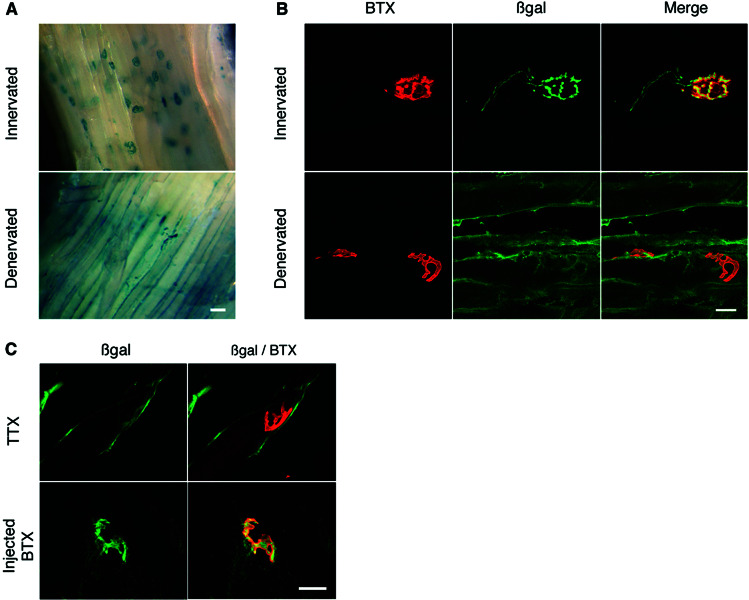
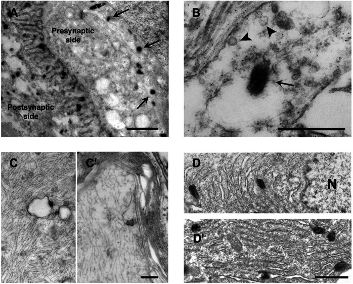
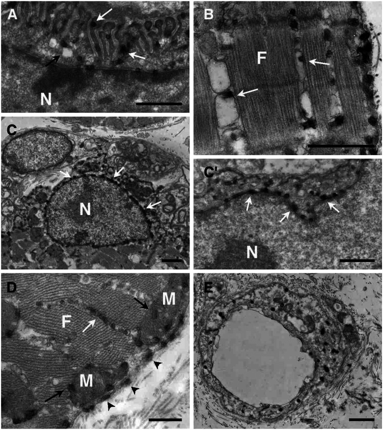
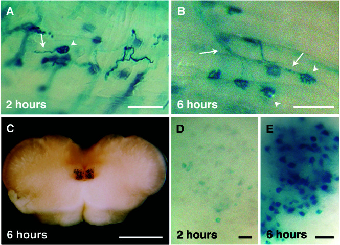
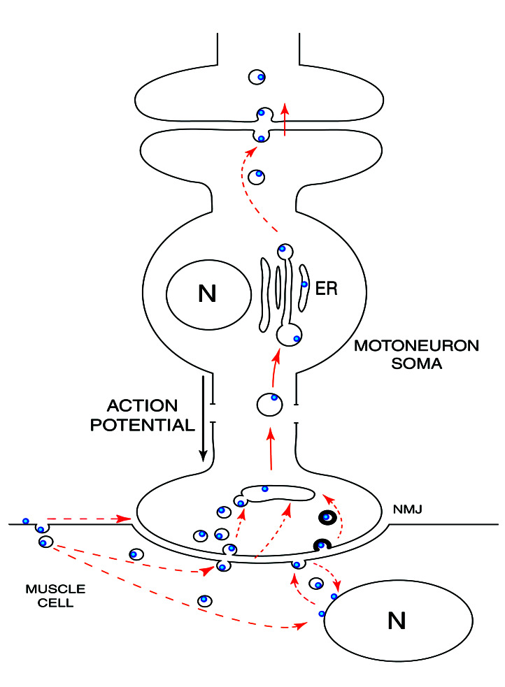
During development and also in adulthood, synaptic connections are modulated by neuronal activity. To follow such modifications in vivo, new genetic tools are designed. The nontoxic C-terminal fragment of tetanus toxin (TTC) fused to a reporter gene such as LacZ retains the retrograde and transsynaptic transport abilities of the holotoxin itself. In this work, the hybrid protein is injected intramuscularly to analyze in vivo the mechanisms of intracellular and transneuronal traffics at the neuromuscular junction (NMJ). Traffic on both sides of the synapse are strongly dependent on presynaptic neural cell activity. In muscle, a directional membrane traffic concentrates beta-galactosidase-TTC hybrid protein into the NMJ postsynaptic side. In neurons, the probe is sorted across the cell to dendrites and subsequently to an interconnected neuron. Such fusion protein, sensitive to presynaptic neuronal activity, would be extremely useful to analyze morphological changes and plasticity at the NMJ.


Figures

Figure 1
Figure 1
Activity-dependent β-gal-TTC localization at the NMJ. (A) The fusion protein was injected intramuscularly in innervated gastrocnemius or 18 h after sciatic nerve transection. Protein localization was investigated 6 h later in toto by X-Gal coloration. β-Gal-TTC protein was localized mainly at the NMJ in innervated gastrocnemius, whereas the denervated muscle showed a diffuse distribution of the fusion protein along the surface structures, with no labeling in the motoneuron being detected. (B) Coimmunocytolocalization of β-gal-TTC by an anti-β-gal antibody and of the NMJ by BTX. In innervated muscle (Upper), a strong β-gal-TTC concentration was detected at the NMJ and into axons. By contrast, in denervated gastrocnemius (Lower), the fusion protein was not concentrated specifically in inactive NMJ. A weak colabeling observed in some cases was not significant as determined by using the Student's t test with a standard statistical threshold of a 5% significance (P < 0.001). (C) Confocal colocalization analysis after TTX blockage of nerve conduction (Upper) or after postsynaptic transmission blockage by BTX (Lower). After TTX administration, the same β-gal-TTC distribution was observed as that in denervated muscle. Injection of a saturating dose of fluorescently labeled BTX, which stops muscle stimulation without affecting nerve functionality, had no effect on β-gal-TTC distribution. (Scale bars: A, 40 μm; B–C, 20 μm.)
Figure 2
Figure 2
β-Gal-TTC neuronal localization by electron microscopy. Electron microscopic analysis was performed by using X-Gal reaction with the electron-dense 5-bromo-4-chloro-3-indol precipitate. (A) On the NMJ motoneuronal side, X-Gal signals (arrows) were detected away from the active zone into large uncoated vesicles (B, arrow); no labeling was detected in small coated synaptic vesicles (B, arrowhead). (C and C′) Staining along the axon was associated with the membranes of large vesicles. (D) In motoneuronal soma, precipitate was found in the perinuclear endoplasmic reticulum. N; nuclei. (Scale bars: A, 1 μm; B, 0.4 μm; C and C′, 0.2 μm; D and D′, 0.5 μm.)
Figure 3
Figure 3
β-Gal-TTC intramuscular localization by electron microscopy. (A) A strong fusion protein concentration was observed on the NMJ postsynaptic side. Staining was detected associated with the junctional folds membranes (white arrows) and vesicular membranes (black arrows). Labeling also was observed in the sarcoplasmic reticulum (white arrows, B and D). Around the subjunctional nuclei, staining was associated with the endoplasmic reticulum (C and C′, white arrows). (D) Precipitate also was observed in mitochondria (black arrows) and associated with the muscle plasma membrane (arrowheads). (E) In blood vessel endothelial cells, an intense staining also was detected, linked to smaller vesicle membranes. F, myofibrils; N, nuclei; M, mitochondria. (Scale bars, 1 μm.)
Figure 4
Figure 4
β-Gal-TTC transport kinetics into the hypoglossal nuclei. The hybrid protein was injected intramuscularly into the tongue, and an in toto X-Gal staining was performed 2 (A and D) or 6 (B, C, and E) h later. β-Gal-TTC was concentrated rapidly at the NMJ and taken up by the connected axons (A and B, arrowheads and arrows, respectively). Motoneuronal soma in the hypoglossal nuclei displayed a weaker intensity 2 h (D) than 6 h after injection (E). (C) Brainstem section showing the hypoglossal nuclei stained 6 h postinjection. (Scale bars: A–B, 100 μm; C, 1 mm; D–E, 20 μm.)
Figure 5
Figure 5
β-Gal-TTC presumed pathways. After intramuscular injection, β-gal-TTC is concentrated rapidly at the NMJ. The dashed arrows represent various hypothetical pathways. In motoneurons, the hybrid protein is found in large uncoated vesicles and then transported retrogradely to the endoplasmic reticulum (ER). Internalization by an interconnected neuron will then occur. N, nuclei.

References

    1. Coen L, Osta R, Maury M, Brûlet P. Proc Natl Acad Sci USA. 1997;94:9400–9405. - PMC - PubMed
    1. Bonnerot C, Rocancourt D, Briand P, Grimber G, Nicolas J F. Proc Natl Acad Sci USA. 1987;84:6795–6799. - PMC - PubMed
    1. Costanzo E M, Barry J A, Ribchester R R. Nat Neurosci. 2000;3:694–700. - PubMed
    1. Jasmin B J, Antony C, Changeux J P, Cartaud J. Eur J Neurosci. 1995;7:470–479. - PubMed
    1. Goldstein L S B, Yang Z. Annu Rev Neurosci. 2000;23:39–71. - PubMed

Publication types

LinkOut - more resources