

Proliferation of T-cell subsets that contact tumour cells in colorectal cancer
- PMID: 11882037
- PMCID: PMC1906282
- DOI: 10.1046/j.1365-2249.2002.01730.x
Proliferation of T-cell subsets that contact tumour cells in colorectal cancer
Abstract
We have investigated the proliferation rates of T-cell subsets in colorectal carcinomas using immunohistochemistry. It was found that the tumour-infiltrating T cells in contact with the tumour cells have a significantly higher frequency of proliferation than those in the stroma. In particular, the CD8+ intraepithelial lymphocytes (T-IEL) within the tumours have a significantly higher frequency of proliferation in comparison with CD8+ T cells in the stromal compartment or in any normal mucosal lymphoid tissues. It is possible that the proliferation of the CD8+ T-IEL may be driven by self-antigens expressed on the tumour cells. The proportion of CD3+ CD7- T cells is increased within carcinomas compared with the normal colon, and a population of CD57+ T cells was observed which is absent from the normal colon. It is possible that these phenotypes are acquired in situ due to repeated stimulation of the T cells by tumour antigens. Intact colorectal carcinoma explants were cultured, and the presence of tumour-infiltrating T cells analysed after 3 days of culture in isolation from the systemic compartments. CD3+ T cells were proliferating (at a low rate) within the explants after 3 days of culture, indicating that they may be sustained by factors present in the tumour microenvironment.
Figures

 ) and colorectal carcinoma-infiltrating stromal cells (□).
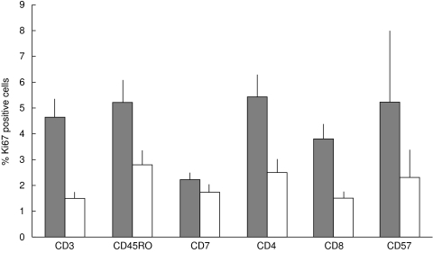 ) and in stromal cells (□).
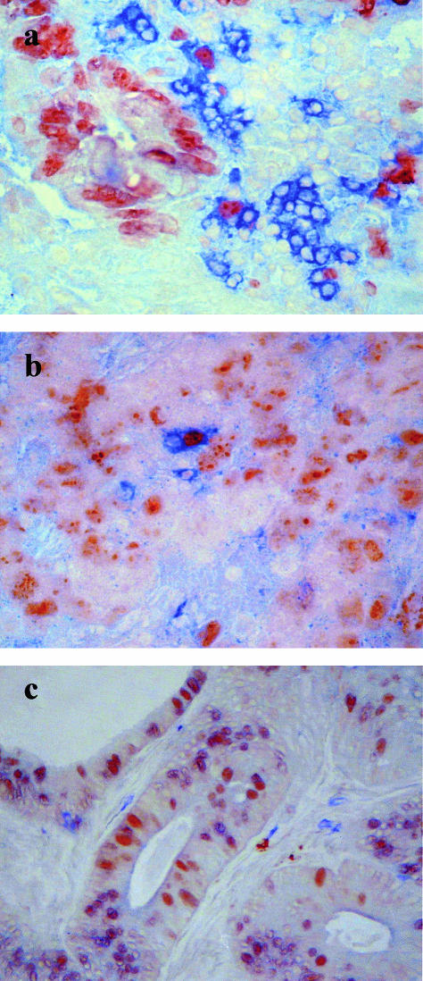
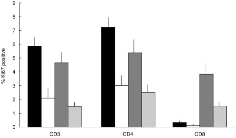 ) and colorectal carcinoma stroma (
 ). The CD8+ subset is proliferative in the carcinoma specimens, but not in the tonsils or the Peyer’s patches.
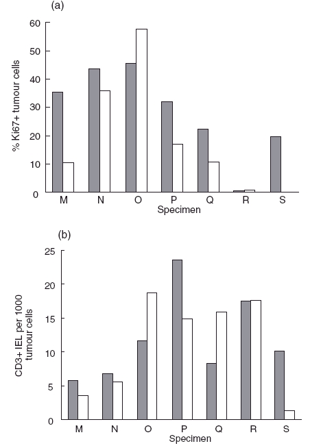 ) and after 3 days of culture (□); (b) the numbers of CD3+ cells in the IEL compartment were determined on day 0 () and day 3 (□) of culture of specimens of colorectal carcinomas.
References
-
- Carlon CA, Fabris G, Arslan-Pagnini C, et al. Prognostic correlations of operable carcinoma of the rectum. Dis Colon Rectum. 1985;28:47–50. - PubMed
-
- Ropponen KM, Eskilinen MJ, Lipponen PK, et al. Prognostic value of tumour-infiltrating lymphocytes (TILs) in colorectal cancer. J Pathol. 1997;182:318–24. 10.1002/(sici)1096-9896(199707)182:3<318::aid-path862>3.0.co;2-6. - DOI - PubMed
Publication types
MeSH terms
Substances
LinkOut - more resources
Full Text Sources
Medical
Research Materials
