. 2002 Mar 1;539(Pt 2):433-44.
doi: 10.1113/jphysiol.2001.013322.

Prostaglandin E(2) inhibits calcium current in two sub-populations of acutely isolated mouse trigeminal sensory neurons

Affiliations

Prostaglandin E(2) inhibits calcium current in two sub-populations of acutely isolated mouse trigeminal sensory neurons

Stephanie L Borgland et al. J Physiol. .

Abstract

Prostaglandins are important mediators of pain and inflammation. We have examined the effects of prostanoids on voltage-activated calcium currents (I(Ca)) in acutely isolated mouse trigeminal sensory neurons, using standard whole cell voltage clamp techniques. Trigeminal neurons were divided into two populations based on the presence (Type 2) or absence (Type 1) of low voltage-activated T-type I(Ca). The absence of T-type I(Ca) is highly correlated with sensitivity to mu-opioid agonists and the VR1 agonist capsaicin. In both populations of cells, high voltage-activated I(Ca) was inhibited by PGE(2) with an EC(50) of about 35 nM, to a maximum of 30 %. T-type I(Ca) was not inhibited by PGE(2). Pertussis toxin pre-treatment abolished the effects of PGE(2) in Type 2 cells, but not in Type 1 cells, whereas treatment with cholera toxin prevented the effects of PGE(2) in Type 1 cells, but not in Type 2 cells. Inhibition of I(Ca) by PGE(2) was associated with slowing of current activation and could be relieved with a large positive pre-pulse, consistent with inhibition of I(Ca) by G protein betagamma subunits. Reverse transcription-polymerase chain reaction of mRNA from trigeminal ganglia indicated that all four EP prostanoid receptors were present. However, in both Type 1 and Type 2 cells the effects of PGE(2) were only mimicked by the selective EP(3) receptor agonist ONO-AE-248, and not by selective agonists for EP(1) (ONO-DI-004), EP(2) (ONO-AE1-259) and EP(4) (ONO-AE1-329) receptors. These data indicate that two populations of neurons in trigeminal ganglia differing in their calcium channel expression, sensitivity to mu-opioids and capsaicin also have divergent mechanisms of PGE(2)-mediated inhibition of calcium channels, with Gi/Go type G proteins involved in one population, and Gs type G proteins in the other.


Figures
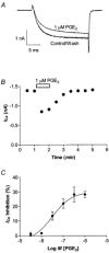
Figure 1
Figure 1. PGE2 inhibits HVA ICa in Type 1 mouse trigeminal neurons
A, HVA ICa elicited by stepping the membrane potential from −80 to 0 mV was inhibited by 1 μm PGE2 in a Type 1 neuron. B, a representative time plot illustrating that inhibition of HVA ICa by 1 μm PGE2 completely reversed upon washout of the drug. C, Type-1 neurons were superfused with various concentrations of PGE2 for 1 min and then washed until the current returned to pre-drug treatment amplitude. Each point represents the mean and s.e.m. of 6–12 cells.
Figure 2
Figure 2. Calcium currents in Type 2 trigeminal neurons are inhibited by PGE2
A, to elicit the low voltage-activated T-type ICa, neurons were stepped from −80 mV to −40 mV. Neurons recovered for 80 ms (approximately 65 ms has been removed for clarity) before stepping the membrane potential to 0 mV to elicit HVA ICa. T-type ICa was not inhibited by 1 μm PGE2. This trace was not leak subtracted and zero current is denoted by the dashed line.B, time course of inhibition of HVA ICa by 1 μm PGE2 in the same cell. C, Type 2 neurons were superfused with various concentrations of PGE2 for 1 min and then washed until the current returned to pre-drug treatment amplitude. Each point represents the mean and s.e.m. of 6–12 cells.
Figure 3
Figure 3. Characteristics of PGE2 modulation of HVA ICa
A and B, HVA ICa was elicited by stepping the membrane potential from −80 mV to potentials between −60 mV and +60 mV in 10 mV increments (Control, black squares). PGE2 (1 μm; triangles) inhibited ICa over a range of membrane potentials. A, a representative example of a current-voltage relationship of a Type 1 cell. B, current-voltage relationship for a Type 2 cell. In Type 1 cells (C) and Type 2 cells (D) 1 μm PGE2 did not alter Cd2+-insensitive current when HVA ICa was elicited by stepping the cells from the holding potential to 0 mV.
Figure 4
Figure 4. Inhibition of HVA ICa by PGE2 is mediated via multiple G proteins
Neurons were treated overnight with either 100 ng ml−1 PTX or 250 ng ml−1 CTX. Aa, CTX selectively abolished PGE2-mediated inhibition of HVA ICa in Type 1 neurons, while PTX treatment selectively reduced effects of PGE2 in Type 2 neurons. Type 2 neurons pre-treated with PTX had significantly less inhibition of HVA ICa than controls (P < 0.001). Ab, a representative trace of a CTX treated Type 1 neuron in the presence and absence of 1 μm PGE2. Ac, a representative trace of a PTX treated Type 2 neuron in the presence and absence of 1 μm PGE2. B, in Type 1 neurons, inhibition of ICa caused by 1 μm ME was abolished by PTX but not CTX. Representative traces of a CTX treated neuron (Bb) and a PTX treated neuron (Bc) in the presence and absence of 1 μm ME. C, ICa inhibition caused by 30 μm baclofen was abolished by PTX, but not CTX in Type 2 cells. Representative traces of a CTX treated Type 2 neuron (Cb) and a PTX treated Type 2 neuron (Cc) in the presence and absence of 30 μm baclofen. Records illustrated are superimposed currents recorded before, during and after drug application.
Figure 5
Figure 5. PGE2 inhibition of HVA ICa in sensory neurons is relieved by a positive prepulse
Different prepulse protocols were used for Type 1 and Type 2 neurons to achieve relief of ICa inhibition. Trigeminal neurons were voltage clamped at −80 mV and were stepped initially (S1) to a test potential of 0 mV. In Type 1 neurons, a 70 ms step to +130 mV was applied. Cells recovered for 20 ms before a second test pulse (S2) was applied. In Type 2 neurons, a 50 ms step to +100 ms was applied. Cells recovered for 15 ms before the second test pulse. A section of approximately 80 ms in the current trace has been omitted for clarity. Inhibition of HVA ICa by 1 μm PGE2 was relieved after a large positive prepulse in both Type 1 and Type 2 cells, but a greater depolarizing pulse was required to achieve relief in Type 1 neurons.
Figure 6
Figure 6. Trigeminal ganglion cells express 4 EP prostanoid receptor subtypes
Total trigeminal ganglion mRNA was extracted and subjected to RT PCR utilizing the primers outlined in Table 1. The resulting PCR products were separated on a 2 % agarose gel stained with ethidium bromide, as illustrated. Lane A is a nucleotide size ladder in 100 bp increments, lanes B, C, D and E contain the amplified DNA fragments corresponding to EP1, EP2, EP3 and EP4 receptors respectively, lane F is the amplified DNA fragment corresponding to hypoxanthine phosphoribosyltransferase, a housekeeping enzyme, lane G contains all the PCR reaction components without added reverse transcription product. This is representative gel from the ganglia of 1 of 13 mice (8 male, 5 female).
Figure 7
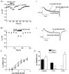
Figure 7. The EP3 agonist ONO-AE-248 inhibits ICa in Type 1 and Type 2 trigeminal neurons
Representative time plots of ONO-AE-248 inhibition of peak ICa in a Type 1 (Aa) and Type 2 (Ba) neuron. ICa was elicited by stepping the membrane potential from −80 to 0 mV. Example traces from the Type 1 neuron (Ab) and Type 2 (Bb) neurons are shown in Aa and Ba. C, the inhibition of ICa by ONO-AE-248 in Type 1 (triangles) and Type 2 (squares) neurons was concentration dependent, with an EC50 of 580 nm and 1.4 μm, respectively. Each point represents the mean and s.e.m. of 6–10 cells. D, overnight treatment with PTX (100 ng ml−1) strongly reduced the ONO-AE-248 (10 μm) inhibition of ICa in Type 2 but not Type 1 cells, whereas treatment with CTX (250 ng ml−1) overnight blocked the ONO-AE-248 inhibition of ICa in Type 1 but not Type 2 cells (n = 6–15 for each).

References

    1. Abdulla FA, Smith PA. Ectopic alpha2-adrenoceptors couple to N-type Ca2+ channels in axotomized rat sensory neurons. Journal of Neuroscience. 1997;17:1633–41. - PMC - PubMed
    1. Arnot MI, Stotz SC, Jarvis SE, Zamponi GW. Differential modulation of N-type α1B and P/Q-type α1A calcium channels by different G protein β subunit isoforms. Journal of Physiology. 2000;527:203–212. - PMC - PubMed
    1. Baccaglini PI, Hogan PG. Some rat sensory neurons in culture express characteristics of differentiated pain sensory cells. Proceedings of the National Academy of Sciences of the USA. 1983;80:594–598. - PMC - PubMed
    1. Borgland SL, Connor M, Christie MJ. Nociceptin inhibits calcium channel currents in a subpopulation of small nociceptive trigeminal ganglion neurons in mouse. Journal of Physiology. 2001;536:35–47. - PMC - PubMed
    1. Bourinet E, Soong TW, Sutton K, Slaymaker S, Mathews E, Monteil A, Zamponi GW, Nargeot J, Snutch TP. Splicing of a1A subunit gene generates phenotypic variants of P- and Q-type calcium channels. Nature Neuroscience. 1999;2:407–421. - PubMed

MeSH terms

LinkOut - more resources