. 2002 Mar 1;539(Pt 2):603-14.
doi: 10.1113/jphysiol.2001.013133.

Feedback inhibition in the inner plexiform layer underlies the surround-mediated responses of AII amacrine cells in the mammalian retina

Affiliations

Feedback inhibition in the inner plexiform layer underlies the surround-mediated responses of AII amacrine cells in the mammalian retina

Béla Völgyi et al. J Physiol. .

Abstract

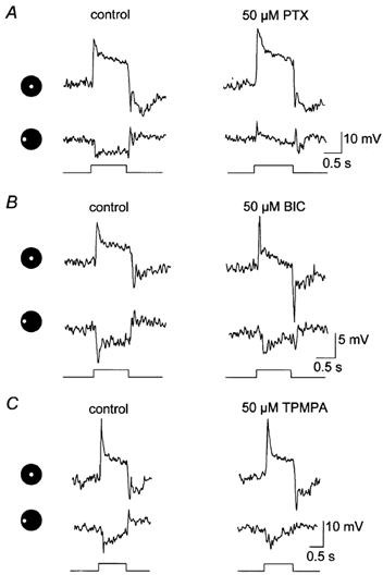
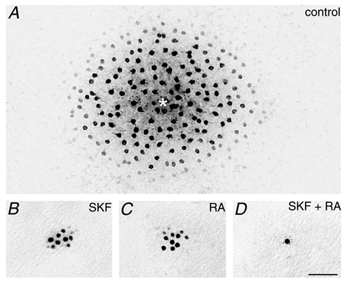
Intracellular recordings were made from narrow-field, bistratified AII amacrine cells in the isolated, superfused retina-eyecup of the rabbit. Pharmacological agents were applied to neurons to dissect the synaptic pathways subserving AII cells so as to determine the circuitry generating their off-surround responses. Application of the GABA antagonists, picrotoxin, bicuculline and 1,2,5,6-tetrahydropyridine-4-yl methylphosphinic acid (TPMPA) all increased the on-centre responses of AII amacrine cells, but attenuated the off-surround activity. At equal concentrations, picrotoxin was approximately twice as effective as bicuculline or TPMPA in modifying the response activity of AII amacrine cells. These results indicate that the mechanism underlying surround inhibition of AII amacrine cells includes activation of both GABA(A) and GABA(C) receptors in an approximately equal ratio. Application of the GABA antagonists also increased the size of on-centre receptive fields of AII amacrine cells. Again, picrotoxin was most effective, producing, on average, a 54 % increase in the size of the receptive field, whereas bicuculline and TPMPA produced comparable 34 and 33 % increases, respectfully. Application of the voltage-gated sodium channel blocker TTX produced effects on AII amacrine cells qualitatively similar to those of the GABA blockers. Intracellular application of the chloride channel blocker 4,4'-dinitro-stilbene-2,2'-disulphonic acid (DNDS) abolished the direct effects of GABA on AII amacrine cells. Moreover, DNDS increased the amplitude of both the on-centre and off-surround responses. The failure of DNDS to block the off-surround activity indicates that it is not mediated by direct GABAergic inhibition. Taken together, our results suggest that surround receptive fields of AII amacrine cells are generated indirectly by the GABAergic, reciprocal feedback synapses from S1/S2 amacrine cells to the axon terminals of rod bipolar cells.


Figures

Figure 1
Figure 1. Effects of GABA blockers on AII amacrine cell light responses
Each panel shows the on-centre and off-surround responses of an AII cell prior to (control) and after a 5 min application of a drug. The icons to the left of each panel and below each response indicate presentation of a 75 μm diameter spot of light either centred over the cell or translated 100 μm peripherally. Stimulus intensity = log −5.5; maximum intensity (log 0.0 = 2.37 mW cm−2). A, effects of 50 μm picrotoxin (PTX) on the light-evoked responses of an AII cell. Picrotoxin enhanced the on-centre response whereas the surround response was abolished revealing a complex depolarizing response. This depolarization is probably a centre-mediated response due to light scatter into the centre-receptive field region. B, application of 50 μm bicuculline (BIC) increased predominantly the transient component of the on-centre response of this cell, but attenuated the off-surround response. C, application of 50 μm TPMPA produced a modest increase in the amplitude of the on-centre response and a reduction in off-surround activity.
Figure 2
Figure 2. Summary of the effects of the GABA blockers on AII cells
A, summary of the effects of the GABA blockers on the amplitude of the on-centre and off-surround responses of AII cells. At equal concentrations of 50 μm, picrotoxin (PTX) was most effective, producing an average 65 % increase in the on-centre response and a 52 % diminution of the off-surround. Bicuculline (BIC) and TPMPA showed similar effects, including an average 27 and 23 % increase in the centre-mediated responses and a 23 and 15 % reduction in the amplitude of the off-surround responses. B, at equal concentrations of 50 μm, PTX produced a 54 % increase in the size of the centre-receptive field of AII cells, whereas BIC and TPMPA only enhanced the receptive field by an average 34 and 33 %, respectively. The standard deviations are represented as a line above each data set. Individual drug treatments (n) = 24 for PTX, 22 for BIC and 23 for TPMPA.
Figure 3
Figure 3. Effects of GABA blockers on the on-centre receptive fields of AII cells
Scatterplots compare the normalized on-centre responses of an AII cell to a 50 μm wide × 6.0 mm long rectangular slit of light presented at different distances from the centre of the cell (0 μm). Values next to each curve indicate diameter (d) of Gaussian functions fitted to the data. A, effect of a 5 min application of 50 μm picrotoxin (PTX) on the centre-receptive field. Prior to application of the drug (control), the cell displayed a centre-receptive field of 219 μm. PTX increased the receptive field by 148 μm or 68 %. B, application of 50 μm bicuculline (BIC) produced a 41 % increase in the centre-receptive field of this AII cell. C, application of 50 μm TPMPA produced a 33 μm increase in the centre-receptive field of this AII cell. Number of drug treatments (n) = 18 for PTX, 19 for BIC and 18 for TPMPA.
Figure 4
Figure 4. Effects of TTX on AII cell responses
A, application of 0.5 μm TTX increased the amplitude of the on-centre response of this AII cell, whereas the off-surround response was completely abolished. Conventions are the same as Fig. 1. B, application of 50 μm TTX increased the size of the centre-receptive field of this AII cell by 83 μm. Conventions are the same as in Fig. 3. C, summary of the effects of TTX on AII cells (n = 16). TTX increased the on-centre responses of AII cells by an average of 41 %, reduced the off-surround responses by 35 % and enlarged the centre-receptive fields by an average of 43 %. Standard deviations are indicated by lines on top of each data set.
Figure 5
Figure 5. Schematic diagram of feedforward and feedback inhibitory circuits subserving AII cells in the IPL
The on-centre receptive field of AII amacrine cells is produced via the rods → rod bipolar cell (RB) → AII amacrine cell pathway. Illumination of the periphery of the AII cell receptive field results in a characteristic off-surround response. There are two possible circuits underlying the surround response. In the first, peripheral signals are carried to the inner retina by the rods →rod bipolar cell → S1-S2 amacrine cell route. In turn, these peripherally generated signals are carried centrally, providing direct GABAergic feedback to rod bipolar cell axon terminals (large circle). Alternatively, an unidentified amacrine cell (?) may provide surround inhibition via direct feedforward inhibitory inputs onto AII amacrine cell dendrites (small circle). Open arrowheads, sign-conserving excitatory synapses; filled arrowheads, converting sign-inverting inhibitory synapses; AII, AII amacrine cell; S1-S2, S1-S2 amacrine cells; OPL, outer plexiform layer; INL, inner nuclear layer; IPL, inner plexiform layer.
Figure 6
Figure 6. Effect of DNDS on GABA-mediated responses of AII cells
A, application of a high Mg2+/low Ca2+ Ringer solution blocked synaptic transmission resulting in a hyperpolarization of the dark membrane potential of the AII cell and a complete blockage of light-evoked on-centre activity. B, during continuous synaptic blockade with the high Mg2+/low Ca2+ solution, application of 250 μm GABA within 1 min of cell impalement produced a 10–15 mV hyperpolarization that was readily reversed following return to the control superfusate. C, a repeated application of GABA 20 min later, after DNDS was allowed to leak into the cell, failed to modulate the dark membrane potential of the cell.
Figure 7
Figure 7. Effects of DNDS on AII cell response activity
Immediately after impalement with a microelectrode loaded with 500 μm DNDS, this AII cell showed a clear on-centre and off-surround responses to a 75 μm diameter spot of light either centred (right) or translated laterally by 100 μm (left). A slight enhancement of the light-evoked responses was detected following 10 min of impalement during which DNDS leaked into the cell. After 20 min, an additional increase in the amplitudes of both the centre- and surround-mediated responses was observed. Application of DNDS also resulted in increased dark background noise and brought out spike activity.
Figure 8
Figure 8. Effects of SKF38393 and retinoic acid on the coupling pattern of AII cells
A, typical tracer-coupled array of AII amacrine cells following injection of Neurobiotin into a single cell (*). Retina was adapted with a dim background light of log −6.0. B, application of the dopamine D1 receptor agonist SKF38393 (400 μm) for 45 min prior to Neurobiotin injection resulted in a large reduction in tracer coupling of AII cells, although some coupling remained. Same adapting conditions as in A. C, application of 150 μm retinoic acid (RA) also reduced the tracer coupling of AII cells from control conditions. D, co-application of both SKF38393 and RA resulted in a complete uncoupling of AII cells so that only the cell injected with Neurobiotin is labelled. Adapting conditions were the same as in A. Calibration bar for all panels = 50 μm.
Figure 9
Figure 9. Effects of DNDS on the light-evoked responses of uncoupled AII cells
Intracellular recordings from the uncoupled AII cell illustrated in Fig. 8D. Immediately after impalement with a microelectrode loaded with 500 μm DNDS, this AII cell showed a clear on-centre and off-surround response to a 75 μm diameter spot of light either centred (right panel) or translated laterally by 100 μm (left panel). Beginning at 10 min after impalement and reaching a maximum effect at 20 min, leakage of DNDS into the cell produced a sizeable increase in both the centre- and surround-mediated responses as well as the dark membrane noise. Therefore, DNDS had the same qualitative effects on both coupled and uncoupled AII amacrine cells.

Similar articles

Cited by

References

    1. Barlow HB, Fitzhugh R, Kuffler SW. Change of organization of receptive fields of the cat's retina during dark adaptation. Journal of Physiology. 1957;137:338–354. - PMC - PubMed
    1. Baylor DA, Fuortes MGF, O'Bryan PM. Receptive field of cones in the retina of turtle. Journal of Physiology. 1971;214:265–294. - PMC - PubMed
    1. Bloomfield SA. Relationship between receptive and dendritic field size of amacrine cells in the rabbit retina. Journal of Neurophysiology. 1992;68:711–725. - PubMed
    1. Bloomfield SA. Effect of spike blockade on the receptive-field size of amacrine and ganglion cells in the rabbit retina. Journal of Neurophysiology. 1996;75:1878–1893. - PubMed
    1. Bloomfield SA, Dacheux RF. Rod vision: pathways and processing in the mammalian retina. Progress in Retina and Eye Research. 2001;20:351–384. - PubMed

Publication types

LinkOut - more resources