

Change in length of relaxed muscle fascicles and tendons with knee and ankle movement in humans
- PMID: 11882694
- PMCID: PMC2290150
- DOI: 10.1113/jphysiol.2001.012756
Change in length of relaxed muscle fascicles and tendons with knee and ankle movement in humans
Abstract
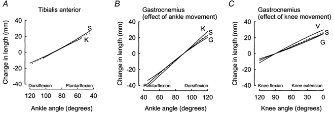
Ultrasonography was used to measure changes in length of muscle fascicles in relaxed human tibialis anterior and gastrocnemius during passively imposed changes in joint angle. Changes in the length of muscle fascicles were compared to changes in the length of the whole muscle-tendon units calculated from joint angles and anthropometric data. Relaxed muscle fascicles underwent much smaller changes in length than their muscle-tendon units. On average, muscle fascicles in tibialis anterior 'saw' 55 +/- 13 % (mean +/- S.D.) of the total change in muscle-tendon length. This indicates nearly half of the total change in muscle-tendon length was taken up by stretch of tendon. In gastrocnemius, which has relatively long tendons, only 27 +/- 9 % of the total change in muscle-tendon length was transmitted to muscle fascicles. Thus, the tendency for passive movement to be taken up by the tendon was greater for gastrocnemius than tibialis anterior (P = 0.002). For these muscles, the relatively large changes in tendon length across much of the physiological range of muscle-tendon lengths could not wholly be explained by tendon slackness, changes in fibre pennation, or stretch or contraction history of the muscle. Our data confirm that when joints are moved passively, length changes 'seen' by muscle fascicles can be much less than changes in the distance between muscle origin and insertion. This occurs because tendons undergo significant changes in length, even at very low forces.
Figures


Similar articles
-
Passive mechanical properties of human gastrocnemius muscle tendon units, muscle fascicles and tendons in vivo.J Exp Biol. 2007 Dec;210(Pt 23):4159-68. doi: 10.1242/jeb.002204. J Exp Biol. 2007. PMID: 18025015 Clinical Trial.
-
Passive elongation of muscle fascicles in human muscles with short and long tendons.Physiol Rep. 2017 Dec;5(23):e13528. doi: 10.14814/phy2.13528. Physiol Rep. 2017. PMID: 29192068 Free PMC article.
-
In vivo motion transmission in the inactive gastrocnemius medialis muscle-tendon unit during ankle and knee joint rotation.J Electromyogr Kinesiol. 2006 Oct;16(5):413-22. doi: 10.1016/j.jelekin.2005.10.001. Epub 2005 Nov 23. J Electromyogr Kinesiol. 2006. PMID: 16309922 Clinical Trial.
-
Biomechanical behavior of muscle-tendon complex during dynamic human movements.J Appl Biomech. 2006 May;22(2):131-47. doi: 10.1123/jab.22.2.131. J Appl Biomech. 2006. PMID: 16871004 Review.
-
Passive changes in muscle length.J Appl Physiol (1985). 2019 May 1;126(5):1445-1453. doi: 10.1152/japplphysiol.00673.2018. Epub 2018 Dec 20. J Appl Physiol (1985). 2019. PMID: 30571291 Review.
Cited by
-
Effects of plyometric training on passive stiffness of gastrocnemii muscles and Achilles tendon.Eur J Appl Physiol. 2012 Aug;112(8):2849-57. doi: 10.1007/s00421-011-2256-x. Epub 2011 Dec 1. Eur J Appl Physiol. 2012. PMID: 22131086 Clinical Trial.
-
The acute effect of stretching on the passive stiffness of the human gastrocnemius muscle tendon unit.J Physiol. 2008 Jan 1;586(1):97-106. doi: 10.1113/jphysiol.2007.140434. Epub 2007 Sep 20. J Physiol. 2008. PMID: 17884924 Free PMC article.
-
The effects of knee joint angle on neuromuscular activity during electrostimulation in healthy older adults.J Rehabil Assist Technol Eng. 2018 Aug 21;5:2055668318779506. doi: 10.1177/2055668318779506. eCollection 2018 Jan-Dec. J Rehabil Assist Technol Eng. 2018. PMID: 31191945 Free PMC article.
-
Architectural anatomy of the human tibialis anterior presents morphological asymmetries between superficial and deep unipennate regions.J Anat. 2023 Oct;243(4):664-673. doi: 10.1111/joa.13864. Epub 2023 Mar 30. J Anat. 2023. PMID: 36999195 Free PMC article.
-
Load and failure behavior of human muscle samples in the context of proximal femur replacement.BMC Musculoskelet Disord. 2016 Apr 6;17:149. doi: 10.1186/s12891-016-0998-7. BMC Musculoskelet Disord. 2016. PMID: 27048598 Free PMC article.
References
-
- Alexander RM, Bennet-Clark HC. Storage of elastic strain energy in muscle and other tissues. Nature. 1977;265:114–117. - PubMed
-
- Armitage P, Berry G. Statistical Methods in Medical Research. 3. Oxford: Blackwell Science; 1994. pp. 288–290.
-
- Binder MD, Stuart DG. Responses of Ia and spindle group II afferents to single motor-unit contractions. Journal of Neurophysiology. 1980;43:621–629. - PubMed
-
- Burgess PR, Wei JY, Clark FJ, Simon JB. Signaling of kinesthetic information by peripheral sensory receptors. Annual Review of Neuroscience. 1982;5:171–187. - PubMed
-
- Cameron WE, Binder MD, Botterman BR, Reinking RM, Stuart DG. “Sensory partitioning” of cat medial gastrocnemius muscle by its muscle spindles and tendon organs. Journal of Neurophysiology. 1983;46:32–47. - PubMed
Publication types
MeSH terms
LinkOut - more resources
Full Text Sources
Other Literature Sources