Physiologic basis for the treatment of epiphrenic diverticulum
- PMID: 11882756
- PMCID: PMC1422440
- DOI: 10.1097/00000658-200203000-00006
Physiologic basis for the treatment of epiphrenic diverticulum
Abstract
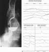
Objective: To quantitate and characterize the motility abnormalities present in patients with epiphrenic diverticula and to assess the outcome of surgical treatment undertaken according to these abnormalities.
Summary background data: The concept that epiphrenic diverticula are complications of esophageal motility disorders rather than primary anatomic abnormalities is gradually becoming accepted. The inconsistency in identifying motility abnormalities in patients with epiphrenic diverticula is a major obstacle to the general acceptance of this concept.
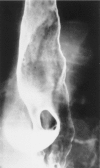
Methods: The study population consisted of 21 consecutive patients with epiphrenic diverticula. All patients underwent videoesophagography, upper gastrointestinal endoscopy, and esophageal motility studies. The diverticula ranged in size from 3 to 10 cm and were predominantly right-sided. Seventeen patients underwent transthoracic diverticulectomy or diverticulopexy with esophageal myotomy and an antireflux procedure. The length of the myotomy was determined by the extent of the motility abnormality. Transhiatal esophagectomy was performed in one patient with multiple diverticula. Two patients declined surgical treatment and another patient died of aspiration before surgery. Symptomatic outcome was assessed via a questionnaire at a median of 24 months after surgery.
Results: The primary symptoms were dysphagia in 5 (24%) patients, dysphagia and regurgitation in 11 (52%) patients, and pulmonary symptoms in 5 (24%) patients. The median duration of the primary symptoms was 10 years. Esophageal motility abnormalities were identified in all patients. An esophageal motor disorder was diagnosed only by 24-hour ambulatory motility testing in one patient, and 24-hour ambulatory motility testing clarified the motility diagnosis in five other patients. The most common underlying disorder was achalasia, which was detected in nine (43%) patients. A hypertensive lower esophageal sphincter was diagnosed in three patients, diffuse esophageal spasm in five, "nutcracker" esophagus in two, and a nonspecific motor disorder in two patients. One patient had an intraoperative myocardial infarction and died. Two patients had persistent mild dysphagia after surgery. The remaining patients had complete relief of their primary symptoms.
Conclusions: There is a high prevalence of named motility disorders in patients with epiphrenic diverticula, and this condition is associated with the potential for lethal aspiration. Twenty-four-hour ambulatory motility testing can be helpful if the results of the stationary examination are normal or indefinite. Resection of the diverticula and a surgical myotomy of the manometrically defined abnormal segment results in relief of symptoms and protection from aspiration.
Figures


References
-
- Mondiere JT. Notes sur quelques maladies de l’oesophage. Arch Gen Med Paris 1833; 3: 28–65.
-
- Rivkin L, Bremner CG, Bremner CH. Pathophysiology of mid-oesophageal and epiphrenic diverticula of the oesophagus. S Afr Med J 1984; 66: 127–129. - PubMed
-
- Streitz JM Jr, Glick ME, Ellis FH, Jr. Selective use of myotomy for treatment of epiphrenic diverticula. Manometric and clinical analysis. Arch Surg 1992; 127: 585–587. - PubMed
-
- Benacci JC, Deschamps C, Trastek VF, et al. Epiphrenic diverticulum: results of surgical treatment. Ann Thorac Surg 1993; 55: 1109–1113. - PubMed
-
- Hudspeth DA, Thorne MT, Conroy R, Pennell TC. Management of epiphrenic esophageal diverticula. A fifteen-year experience. Am Surgeon 1993; 59: 40–42. - PubMed
MeSH terms
LinkOut - more resources
Full Text Sources

