Comparative Study
. 2002 Apr;76(7):3267-75.
doi: 10.1128/jvi.76.7.3267-3275.2002.

The fusion peptide of Semliki Forest virus associates with sterol-rich membrane domains

Affiliations
Comparative Study

The fusion peptide of Semliki Forest virus associates with sterol-rich membrane domains

Anna Ahn et al. J Virol. 2002 Apr.

Abstract

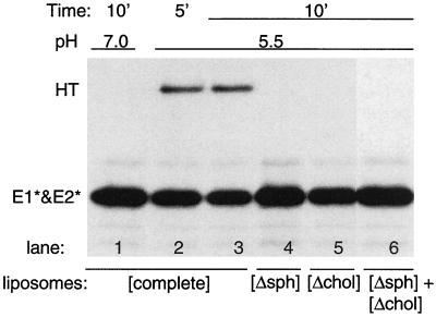
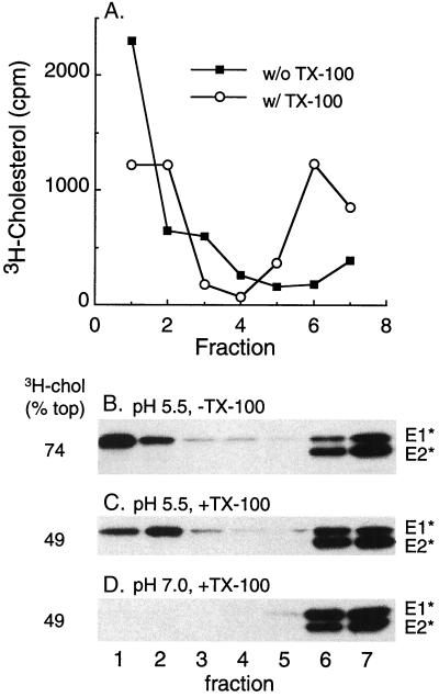
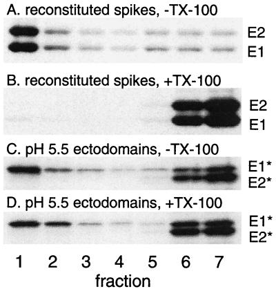
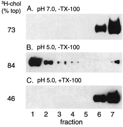
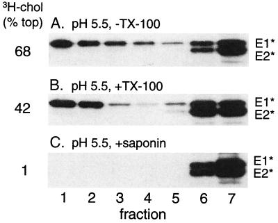
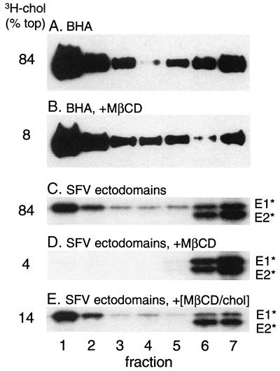
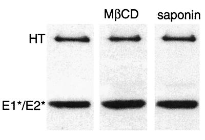
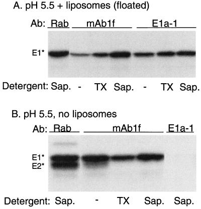
Semliki Forest virus (SFV) is an enveloped alphavirus whose membrane fusion is triggered by low pH and promoted by cholesterol and sphingolipid in the target membrane. Fusion is mediated by E1, a viral membrane protein containing the putative fusion peptide. Virus mutant studies indicate that SFV's cholesterol dependence is controlled by regions of E1 outside of the fusion peptide. Both E1 and E1*, a soluble ectodomain form of E1, interact with membranes in a reaction dependent on low pH, cholesterol, and sphingolipid and form highly stable homotrimers. Here we have used detergent extraction and gradient floatation experiments to demonstrate that E1* associated selectively with detergent-resistant membrane domains (DRMs or rafts). In contrast, reconstituted full-length E1 protein or influenza virus fusion peptide was not associated with DRMs. Methyl beta-cyclodextrin quantitatively extracted both cholesterol and E1* from membranes in the absence of detergent, suggesting a strong association of E1* with sterol. Monoclonal antibody studies demonstrated that raft association was mediated by the proposed E1 fusion peptide. Thus, although other regions of E1 are implicated in the control of virus cholesterol dependence, once the SFV fusion peptide inserts in the target membrane it has a high affinity for membrane domains enriched in cholesterol and sphingolipid.


Figures

FIG. 1.
FIG. 1.
Lipid requirements for formation of the E1* homotrimer. [35S]methionine-labeled ectodomains were mixed with various types of liposomes at a final lipid concentration of 1 mM. The liposomes were either “complete liposomes” containing PE, PC, cholesterol, and sphingomyelin ([complete]) or were deficient in either sphingomyelin ([Δsph]) or cholesterol ([Δchol]). One sample ([Δsph] + [Δchol]) contained both deficient liposome types, each at a lipid concentration of 1 mM. All samples were preincubated for 5 min at 37°C and then treated at the indicated pH at 37°C for the indicated time. The samples were then solubilized in SDS gel buffer at 30°C in the absence of reduction and alkylation, conditions that preserve the E1* homotrimer (HT) but do not separate E1* and E2*, and analyzed by SDS-PAGE. A representative example of two experiments is shown.
FIG. 2.
FIG. 2.
Membrane-inserted E1* is associated with DRM domains. (A) Sucrose gradient floatation pattern of [3H]cholesterol-labeled liposomes before and after extraction with TX-100. Complete liposomes containing trace amounts of [3H]cholesterol were extracted on ice for 10 min with 1% TX-100 where indicated and were loaded on the bottom of a sucrose step gradient. The gradients were centrifuged to float the liposomes to the top of the gradient and were fractionated, and the radioactive cholesterol was quantified. Fraction 1 is the top of the gradient. (B to D) [35S]methionine-labeled ectodomains were mixed with complete liposomes at a final lipid concentration of 1 mM, treated at the indicated pH for 10 min at 37°C as for Fig. 1, and then adjusted to neutral pH. The samples were extracted with Triton X-100 on ice where indicated and separated by sucrose gradient floatation as for panel A. The fractions were analyzed by SDS-PAGE and scintillation counting to quantify the position of the viral ectodomains and the [3H]cholesterol, respectively. The percentage of the total [3H]cholesterol recovered in the top three fractions is shown on the left side of the figure and is an average of that obtained for six experiments.
FIG. 3.
FIG. 3.
The TM domains of reconstituted viral E1 and E2 do not associate with DRMs. The DRM association of the E1 ectodomain and of the full-length SFV E1 and E2 proteins was compared. Radiolabeled full-length SFV E1 and E2 were reconstituted into complete liposomes at neutral pH via detergent dialysis (A and B). Alternatively, radiolabeled ectodomains were mixed with complete liposomes previously prepared by detergent dialysis and treated at pH 5.5 for 10 min at 37°C to trigger insertion (C and D). Samples were then extracted with TX-100 on ice where indicated, separated by sucrose gradient floatation, and analyzed as described for Fig. 2. A representative example of two experiments is shown.
FIG. 4.
FIG. 4.
The influenza HA fusion peptide does not associate with DRMs. BHA was mixed with complete liposomes (1 μg of BHA plus 1 mM lipid), treated for 5 min at the indicated pH, returned to neutral pH, and extracted as indicated with TX-100 on ice. The samples were separated by sucrose gradient floatation and analyzed as described for Fig. 2, except that BHA was detected by immunoblotting (see Materials and Methods). A representative example of two experiments is shown. A pH 7 sample was also extracted with TX-100 and showed identical results to those shown in panel A (data not shown).
FIG. 5.
FIG. 5.
Extraction of membrane [3H]cholesterol and E1* with saponin. Radiolabeled ectodomains were mixed with complete liposomes, treated at pH 5.5 for 10 min at 37°C, extracted for 20 min on ice as indicated with either 1% TX-100 or 1% saponin, separated by sucrose gradient floatation, and analyzed as described for Fig. 2. A representative example of two experiments is shown.
FIG. 6.
FIG. 6.
Extraction of membrane [3H]cholesterol, BHA, and E1* with MβCD. BHA or SFV ectodomains were mixed with complete liposomes at a final lipid concentration of 1 mM and treated at low pH for 10 min at 37°C to trigger BHA or E1* membrane binding. Samples were adjusted to neutral pH and then treated where indicated for 30 min at 37°C with either 20 mM MβCD or 20 mM MβCD precomplexed with cholesterol (MβCD/chol). Samples were then separated by sucrose gradient floatation and analyzed as described for Fig. 4. A representative example of two experiments is shown.
FIG. 7.
FIG. 7.
Extracted E1* is a homotrimer. Radiolabeled ectodomains were mixed with complete liposomes at a final lipid concentration of 1 mM, treated at pH 5.5 for 10 min at 37°C as described for Fig. 1, and then adjusted to neutral pH. The samples were extracted as indicated with 20 mM MβCD for 30 min at 37°C or with 1% saponin for 20 min on ice. The samples were then solubilized in nonreducing SDS gel buffer at 30°C and the presence of the E1 homotrimer (HT) was analyzed by SDS-PAGE as described for Fig. 1. A representative example of two experiments is shown.
FIG. 8.
FIG. 8.
E1* associates with rafts via its fusion peptide. Radiolabeled ectodomains were mixed with 1 mM complete liposomes (A) or buffer (B), and the mixtures were acid treated at pH 5.5 for 3 min at 37°C. The sample for panel A was then floated on a sucrose gradient as described for Fig. 2, and the top four fractions containing liposomes and associated protein were collected and pooled. Aliquots of the panel A and B samples were then, where indicated, extracted for 30 min with either 1% TX-100 on ice or 1% saponin at room temperature. The samples were then immunoprecipitated in the absence of any additional detergent using a polyclonal rabbit antiserum against the SFV envelope proteins (Rab), MAb 1f against E1 residues 85 to 95, or MAb E1a-1 against the acid conformation of E1. A representative example of three experiments is shown. −, no detergent.

Similar articles

Cited by

References

    1. Ahmed, S. N., D. A. Brown, and E. London. 1997. On the origin of sphingolipid/cholesterol-rich detergent-insoluble cell membranes: physiological concentrations of cholesterol and sphingolipid induce formation of a detergent-insoluble, liquid-ordered lipid phase in model membranes. Biochemistry 36:10944-10953. - PubMed
    1. Ahn, A., M. R. Klimjack, P. K. Chatterjee, and M. Kielian. 1999. An epitope of the Semliki Forest virus fusion protein exposed during virus-membrane fusion. J. Virol. 73:10029-10039. - PMC - PubMed
    1. Anderson, R. G. W. 1998. The caveolae membrane system. Annu. Rev. Biochem. 67:199-225. - PubMed
    1. Brown, D. A., and E. London. 1998. Functions of lipid rafts in biological membranes. Annu. Rev. Cell Dev. Biol. 14:111-136. - PubMed
    1. Brown, D. A., and E. London. 2000. Structure and function of sphingolipid- and cholesterol-rich membrane rafts. J. Biol. Chem. 275:17221-17224. - PubMed

Publication types

LinkOut - more resources