. 2002 Apr;76(7):3282-91.
doi: 10.1128/jvi.76.7.3282-3291.2002.

Herpes simplex virus tegument protein US11 interacts with conventional kinesin heavy chain

Affiliations

Herpes simplex virus tegument protein US11 interacts with conventional kinesin heavy chain

Russell J Diefenbach et al. J Virol. 2002 Apr.

Abstract

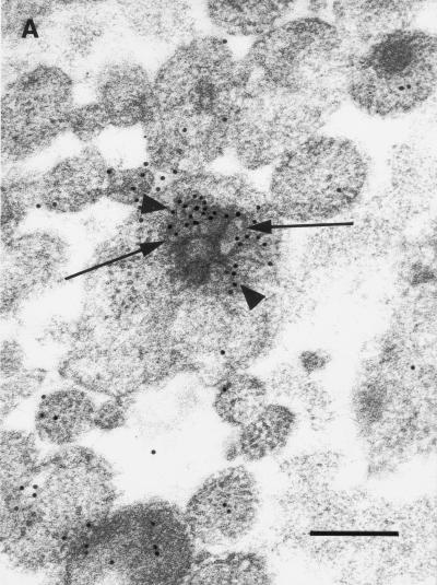
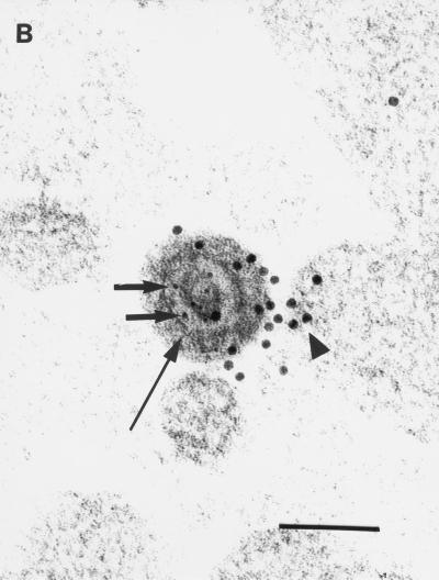
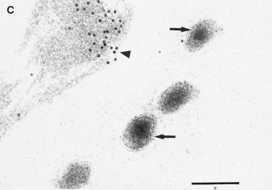
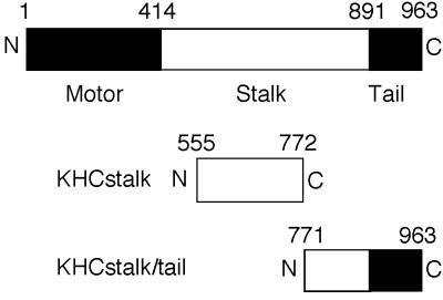
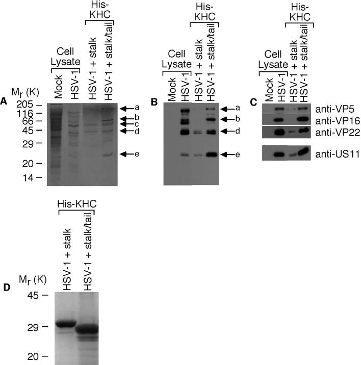
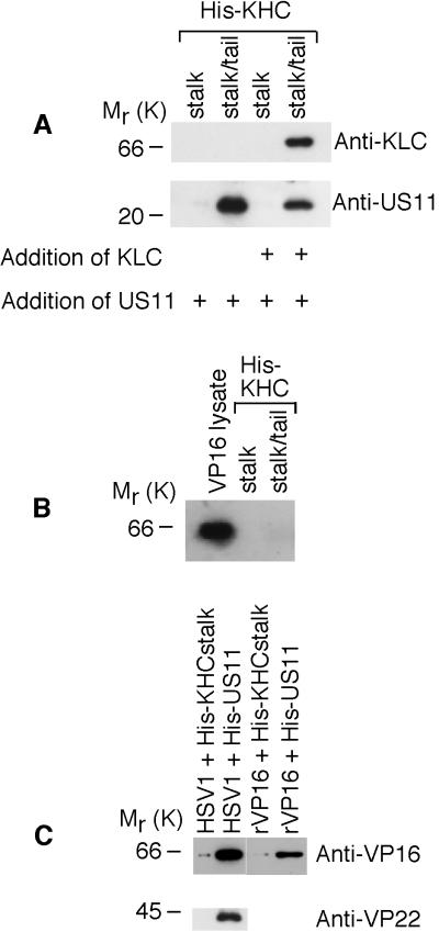
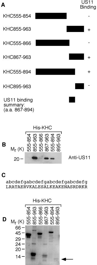
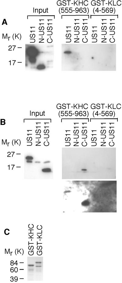
Little is known about the mechanisms of transport of neurotropic herpesviruses, such as herpes simplex virus (HSV), varicella-zoster virus, and pseudorabies virus, within neurons. For these viruses, which replicate in the nucleus, anterograde transport from the cell body of dorsal root ganglion (DRG) neurons to the axon terminus occurs over long distances. In the case of HSV, unenveloped nucleocapsids in human DRG neurons cocultured with autologous skin were observed by immunoelectron microscopy to colocalize with conventional ubiquitous kinesin, a microtubule-dependent motor protein, in the cell body and axon during anterograde axonal transport. Subsequently, four candidate kinesin-binding structural HSV proteins were identified (VP5, VP16, VP22, and US11) using oligohistidine-tagged human ubiquitous kinesin heavy chain (uKHC) as bait. Of these viral proteins, a direct interaction between uKHC and US11 was identified. In vitro studies identified residues 867 to 894 as the US11-binding site in uKHC located within the proposed heptad repeat cargo-binding domain of uKHC. In addition, the uKHC-binding site in US11 maps to the C-terminal RNA-binding domain. US11 is consistently cotransported with kinetics similar to those of the capsid protein VP5 into the axons of dissociated rat neurons, unlike the other tegument proteins VP16 and VP22. These observations suggest a major role for the uKHC-US11 interaction in anterograde transport of unenveloped HSV nucleocapsids in axons.


Figures

FIG. 1.
FIG. 1.
Transmission immunoelectron micrographs of human fetal axons labeled with 5- and 10-nm-diameter immunogold particles recognizing HSV major capsid protein VP5 and the stalk of human uKHC, respectively. Short arrows, 5-nm-diameter gold particles; arrowheads, 10-nm-diameter gold particles. Bars, 200 nm (A and C) and 100 nm (B). (A) Clustering of immunogold label for uKHC around axonal vesicles (long arrows) and diffuse labeling of other axons. Labeling with preimmune sera produced only sparse background label (data not shown). (B) Colocalization of immunogold labels against uKHC and VP5 over a dense unenveloped viral particle in an axon. The long arrow indicates the position of an axonlema. (C) Extracellullar virus labeled only with anti-VP5. In the upper left corner is an axon with diffuse 10-nm immunolabel against uKHC. There is no immunolabel for uKHC over extracellular virus, with only sparse background label outside the axons.
FIG. 1.
FIG. 1.
Transmission immunoelectron micrographs of human fetal axons labeled with 5- and 10-nm-diameter immunogold particles recognizing HSV major capsid protein VP5 and the stalk of human uKHC, respectively. Short arrows, 5-nm-diameter gold particles; arrowheads, 10-nm-diameter gold particles. Bars, 200 nm (A and C) and 100 nm (B). (A) Clustering of immunogold label for uKHC around axonal vesicles (long arrows) and diffuse labeling of other axons. Labeling with preimmune sera produced only sparse background label (data not shown). (B) Colocalization of immunogold labels against uKHC and VP5 over a dense unenveloped viral particle in an axon. The long arrow indicates the position of an axonlema. (C) Extracellullar virus labeled only with anti-VP5. In the upper left corner is an axon with diffuse 10-nm immunolabel against uKHC. There is no immunolabel for uKHC over extracellular virus, with only sparse background label outside the axons.
FIG. 2.
FIG. 2.
Summary of KHC structure and fusion proteins expressed in bacteria. Diagrams of the domain structure of uKHC and fragments of uKHC expressed in bacteria with oligohistidine N-terminal tags are shown. Solid and open areas of the bars delineate protein domains.
FIG. 3.
FIG. 3.
Identification of kinesin-binding HSV-1 proteins. 35S-labeled HSV-1-infected HEp-2 cell lysates were incubated with oligohistidine-tagged uKHC fragments bound to nickel-activated beads. Eluted kinesin-viral-protein complexes were separated by SDS-14% PAGE, transferred to nitrocellulose, and analyzed by autoradiography and immunoblotting. (A) Detection by autoradiography of five 35S-labeled proteins, designated a to e, bound to KHCstalk/tail. (B) Detection with polyclonal antibody to HSV-1 identified bands a, b, d, and e as viral proteins preferentially bound to KHCstalk/tail. (C) The viral protein bands were identified with specific antibodies as the structural proteins VP5 (a), VP16 (b), VP22 (d), and US11 (e). In each immunoblot, mock-infected and HSV-1-infected HEp-2 cell lysates are shown to illustrate antibody specificity. (D) Detection of oligohistidine-tagged KHCstalk and KHCstalk/tail fragments in eluted kinesin-viral-protein complexes. Samples were separated by SDS-12% PAGE and stained with Coomassie blue.
FIG. 4.
FIG. 4.
Interaction of US11 with uKHC and with VP5, VP16, and VP22. Shown are immunoblots of protein complexes eluted from nickel-activated beads and run on SDS-14% PAGE. (A) Lysates of bacteria expressing untagged KLC or US11 were incubated, as indicated (+), with KHCstalk and KHCstalk/tail. US11 bound only to KHCstalk/tail in the absence of other HSV-1 proteins (lower blot). Overnight preincubation of the uKHC fragments with KLC, which binds only to KHCstalk/tail (upper blot), prior to addition of US11 also showed binding of US11 (lower blot). (B) In a similar experiment, recombinant untagged VP16 did not bind to either His-KHC fragment. Expression of VP16 in bacterial lysates was confirmed. (C) Unlabeled HSV-1-infected HEp-2 cell lysate was incubated with KHCstalk and oligohistidine-tagged US11. The viral proteins VP16 and VP22 coeluted with US11 only. Recombinant untagged VP16 was incubated with His-US11 or His-KHCstalk. Recombinant VP16 coeluted with His-US11 only.
FIG.5.
FIG.5.
Identification of the US11-binding site in uKHC. (A) Summary of US11 binding to fragments of uKHC expressed in bacteria with an oligohistidine N-terminal tag. a.a., amino acids. (B) Immunoblot of protein complexes eluted from nickel-activated beads and run on SDS-14% PAGE. Detection was with mouse monoclonal anti-US11. (C) Amino acid sequence of US11-binding site in human uKHC (residues 867 to 894) with repeating heptad motif labeled a to g.The presence of heptad repeats was determined using the COILS server (http://www.ch.embnet.org/software/COILS_form.html), which employs the algorithm of Lupas et al. (19). (D) Eluates from the blot in panel B were run on SDS-14% PAGE and stained with Coomassie blue to detect His-KHC fragments. The position of KHC895-963 is indicated by an arrow.
FIG. 6.
FIG. 6.
Identification of the uKHC-binding site in US11. Protein complexes were eluted from glutathione-Sepharose beads and run on SDS-14% PAGE. (A) Immunoblotting with mouse monoclonal anti-US11 detected full-length US11 bound only to GST-KHC. (B) Immunoblotting with rabbit polyclonal anti-US11 detected only C-US11 bound to GST-KHC (upper blot). Overexposure of the blot also detects full-length US11 bound to GST-KHC (lower blot). (C) Eluates from glutathione-Sepharose beads were run on SDS-14% PAGE and stained with Coomassie blue to detect GST fusion proteins.
FIG. 7.
FIG. 7.
Transport of structural HSV antigens into axons of infected dissociated rat neonatal neurons in vitro. Dissociated DRG neurons were infected with wt HSV-1 (CW1) at 10 PFU/neuron in the absence (A to C) or presence (D and E) of BFA. The appearance and transport into the principal axon of HSV-1 tegument (US11) and nucleocapsid (VP5) antigens was followed by serial fixation over 6 to 24 h postinfection, and then immunofluorescence and confocal microscopy were performed as previously described (23). US11 was present in axons at 24 h (A) but not at 10 h (B). VP5 was also present in axons at 24 h (C) but not at 10 h (reference and data not shown). After treatment of neurons with BFA, US11 (D), but not VP16 (E), was present at 24 h in the majority of axons. Bars, 10 (A and E), 50 (B), and 25 (C and D) μm.

References

    1. Bloom, G. S., M. C. Wagner, K. K. Pfister, and S. T. Brady. 1988. Native structure and physical properties of bovine brain kinesin and identification of the ATP-binding subunit polypeptide. Biochemistry 27:3409-3416. - PubMed
    1. Brideau, A. D., J. P. Card, and L. W. Enquist. 2000. Role of pseudorabies virus Us9, a type II membrane protein, in infection of tissue culture cells and the rat nervous system. J. Virol. 74:834-845. - PMC - PubMed
    1. Brideau, A. D., M. G. Eldridge, and L. W. Enquist. 2000. Directional trans-neuronal infection by pseudorabies virus is dependent on an acidic internalization motif in the Us9 cytoplasmic tail. J. Virol. 74:4549-4561. - PMC - PubMed
    1. Cassady, K. A., M. Gross, and B. Roizman. 1998. The herpes simplex virus US11 protein effectively compensates for the γ134.5 gene if present before activation of protein kinase R by precluding its phosphorylation and that of the α subunit of eukaryotic translation initiation factor 2. J. Virol. 72:8620-8626. - PMC - PubMed
    1. Cohen, G. H., M. Ponce de Leon, H. Diggelmann, W. C. Lawrence, S. K. Vernon, and R. J. Eisenberg. 1980. Structural analysis of the capsid polypeptides of herpes simplex virus types 1 and 2. J. Virol. 34:521-531. - PMC - PubMed

Publication types

LinkOut - more resources