. 2002 Apr;76(7):3365-73.
doi: 10.1128/jvi.76.7.3365-3373.2002.

Coxsackievirus B3 replication is reduced by inhibition of the extracellular signal-regulated kinase (ERK) signaling pathway

Affiliations

Coxsackievirus B3 replication is reduced by inhibition of the extracellular signal-regulated kinase (ERK) signaling pathway

Honglin Luo et al. J Virol. 2002 Apr.

Abstract

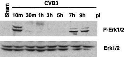
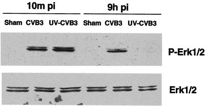
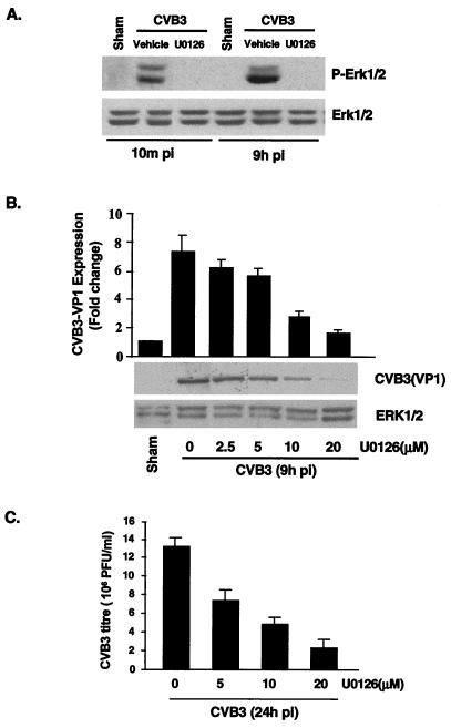
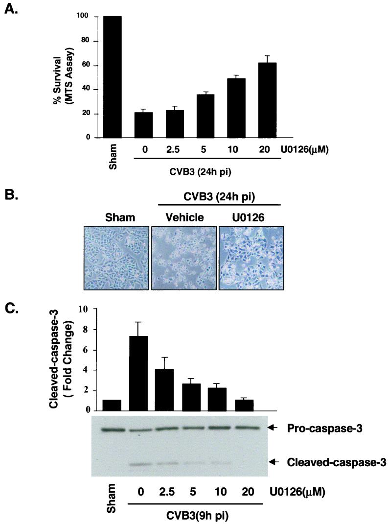
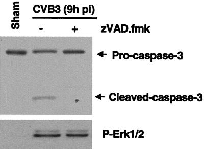
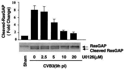
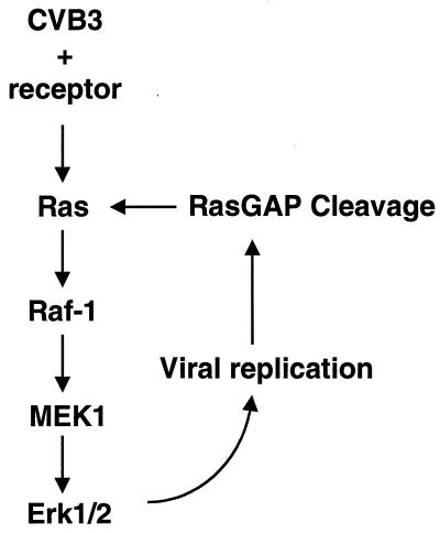
Coxsackievirus B3 (CVB3) is the most common human pathogen for viral myocarditis. We have previously shown that the signaling protein p21(ras) GTPase-activating protein (RasGAP) is cleaved and that mitogen-activated protein kinases (MAPKs) ERK1/2 are activated in the late phase of CVB3 infection. However, the role of intracellular signaling pathways in CVB3-mediated myocarditis and the relative advantages of such pathways to host or virus remain largely unclear. In this study we extended our prior studies by examining the interaction between CVB3 replication and intracellular signaling pathways in HeLa cells. We observed that CVB3 infection induced a biphasic activation of ERK1/2, early transient activation versus late sustained activation, which were regulated by different mechanisms. Infection by UV-irradiated, inactivated virus capable of receptor binding and endocytosis triggered early ERK1/2 activation, but was insufficient to trigger late ERK1/2 activation. By using a general caspase inhibitor (zVAD.fmk) we further demonstrated that late ERK1/2 activation was not a result of CVB3-mediated caspase cleavage. Treatment of cells with U0126, a selective inhibitor of MAPK kinase (MEK), significantly inhibited CVB3 progeny release and decreased virus protein production. Furthermore, inhibition of ERK1/2 activation circumvented CVB3-induced apoptosis and viral protease-mediated RasGAP cleavage. Taken together, these data suggest that ERK1/2 activation is important for CVB3 replication and contributes to virus-mediated changes in host cells. Our findings demonstrate coxsackievirus takeover of a particular host signaling mechanism and uncover a prospective approach to stymie virus spread and preserve myocardial integrity.


Figures

FIG. 1.
FIG. 1.
Time course for CVB3 stimulation and ERK1/2 phosphorylation. Growth-arrested HeLa cells were incubated with CVB3 at an MOI of 10 for 1 h, and then the cells were washed with PBS twice and replenished with serum-free medium. Cell lysates were collected for the indicated times following CVB3 infection (pi) and subjected to Western blotting. ERK1/2 activities were analyzed based on phosphorylated ERK1/2 (P-Erk1/2). To verify equal loading, Western blotting was performed with an antibody to ERK1/2. Data are from one of three different experiments. Sham, sham treatment with PBS for 1 h.
FIG. 2.
FIG. 2.
UV-irradiated CVB3 stimulates early-phase phosphorylation of ERK1/2. HeLa cells were infected with either wild-type virus or UV-irradiated virus, and 10 min and 9 h after infection (pi) cell lysates were harvested and Western blotting was performed to determine ERK activation. To verify equal loading, Western blotting was performed with an anti-ERK1/2 antibody. Data are from one of two different experiments. Sham is as defined for Fig. 1.
FIG. 3.
FIG. 3.
MEK inhibitor reduces viral progeny release and viral protein synthesis. (A) Inhibition of ERK activation by MEK inhibitor U0126 was determined by Western blotting with an anti-phosphorylated ERK antibody. HeLa cells were preincubated with U0126 (20 μM) for 30 min and then were infected with CVB3 (MOI = 10). One hour later, cells were washed twice with PBS and replenished with serum-free medium containing fresh U0126. To verify equal loading, Western blotting was performed with an anti-ERK1/2 antibody. The data are representative of two different experiments. Sham, pi, and P-Erk1/2 are as defined for Fig. 1. (B) HeLa cells were treated with U0126 exactly as for panel A. Cellular lysates were collected from CVB3-infected HeLa cells 9 h postinfection, and Western blot analysis using a CVB3 polyclonal antibody that recognizes viral structure protein VP1 was performed. Results (means ± standard errors [SE]; n = 3) were quantitated by densitometric analysis using National Institutes of Health Image, version 1.61, and normalized to control levels (sham-infected cells without U0126) arbitrarily set to 1.0. (C) HeLa cells were treated with different concentrations of U0126 exactly as for panel A. Medium was collected from CVB3-infected HeLa cells 24 h after infection, and virus titers were determined by plaque assays on HeLa cell monolayers. Values are means ± SE from three independent experiments, in each of which titrations were carried out in triplicate.
FIG. 4.
FIG. 4.
MEK inhibitor blocks CVB3-induced CPE and apoptosis. (A) HeLa cells were treated with U0126 as described for Fig. 3A. Cell viability was determined at 24 h postinfection (pi) by the MTS assay, which measures mitochondrial function, at 24 h after infection. Values are means ± standard errors (SE; n = 6). The level of MTS in sham-infected cells in the absence of U0126 was defined as 100% survival. Similar results were obtained in three independent experiments. (B) Representative phase-contrast microscopy of HeLa cells treated with medium containing or lacking U0126 24 h postinfection. (C) HeLa cells were pretreated with vehicle or various concentrations of U0126 for 30 min, followed by infection with CVB3 for 1 h, 9 h after infection. Western blotting was performed to examine the cleavage of caspase 3. Results were quantitated by densitometric analysis using National Institutes of Health Image, version 1.61, and normalized to the control level (sham-infected cells without U0126), which was arbitrarily set to 1.0. Values are means ± SE (n = 3).
FIG. 5.
FIG. 5.
ERK1/2 activation was not due to caspase activation. HeLa cells were preincubated with zVAD.fmk (100 μM) for 30 min and then infected with CVB3 for 1 h. Cell lysates were collected 9 h postinfection (pi). ERK1/2 activation and caspase 3 cleavage were determined by Western blotting using a phosphorylated ERK1/2 (P-Erk1/2) antibody and a caspase 3 antibody. The data are representative of two different experiments.
FIG. 6.
FIG. 6.
MEK inhibitor inhibits CVB3-mediated RasGAP cleavage. HeLa cells were pretreated for 30 min with various concentrations of U0126 and then were infected by CVB3 for 1 h. At 9 h after addition of CVB3, HeLa cells were harvested and Western blot analysis was performed using an antibody that recognizes RasGAP. Results were quantitated by densitometric analysis using National Institutes of Health Image, version 1.61, and normalized to the control level (sham-infected cells without U0126), which was arbitrarily set to 1.0. Values are means ± standard errors (n = 3).
FIG. 7.
FIG. 7.
A proposed model of the mechanism of ERK activation during CVB3 infection. CVB3 binds with its receptor and initiates early transient ERK phosphorylation. Virus replication mediates RasGAP cleavage, which triggers late-phase ERK phosphorylation. Subsequently, there is a positive feedback to augment ERK activation and viral replication.

Similar articles

Cited by

References

    1. Abe, M. K., W. L. Kuo, M. B. Hershenson, and M. R. Rosner. 1999. Extracellular signal-regulated kinase 7 (ERK7), a novel ERK with a C-terminal domain that regulates its activity, its cellular localization, and cell growth. Mol. Cell. Biol. 19:1301-1312. - PMC - PubMed
    1. Alcorn, M. J., J. L. Booth, K. M. Coggeshall, and J. P. Metcalf. 2001. Adenovirus type 7 induces interleukin-8 production via activation of extracellular regulated kinase 1/2. J. Virol. 75:6450-6459. - PMC - PubMed
    1. Anderson, D. R., J. E. Wilson, C. M. Carthy, D. Yang, R. Kandolf, and B. M. McManus. 1996. Direct interactions of coxsackievirus B3 with immune cells in the splenic compartment of mice susceptible or resistant to myocarditis. J. Virol. 70:4632-4645. - PMC - PubMed
    1. Badorff, C., G. H. Lee, B. J. Lamphear, M. E. Martone, K. P. Campbell, R. E. Rhoads, and K. U. Knowlton. 1999. Enteroviral protease 2A cleaves dystrophin: evidence of cytoskeletal disruption in an acquired cardiomyopathy. Nat. Med. 5:320-326. - PubMed
    1. Beck, M. A., N. M. Chapman, B. M. McManus, J. C. Mullican, and S. Tracy. 1990. Secondary enterovirus infection in the murine model of myocarditis. Pathologic and immunologic aspects. Am. J. Pathol. 136:669-681. - PMC - PubMed

Publication types

MeSH terms