Comparative Study
. 2002 Apr;76(7):3482-92.
doi: 10.1128/jvi.76.7.3482-3492.2002.

Structural analysis of the hepatitis C virus RNA polymerase in complex with ribonucleotides

Affiliations
Comparative Study

Structural analysis of the hepatitis C virus RNA polymerase in complex with ribonucleotides

Stéphane Bressanelli et al. J Virol. 2002 Apr.

Abstract

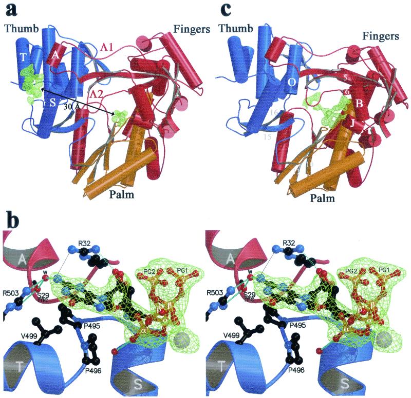
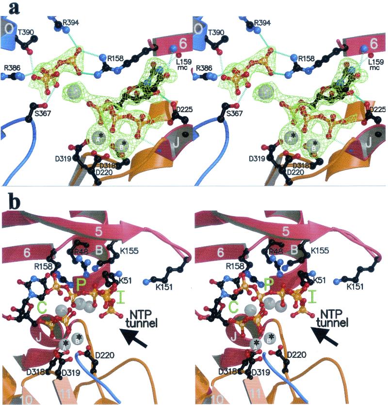
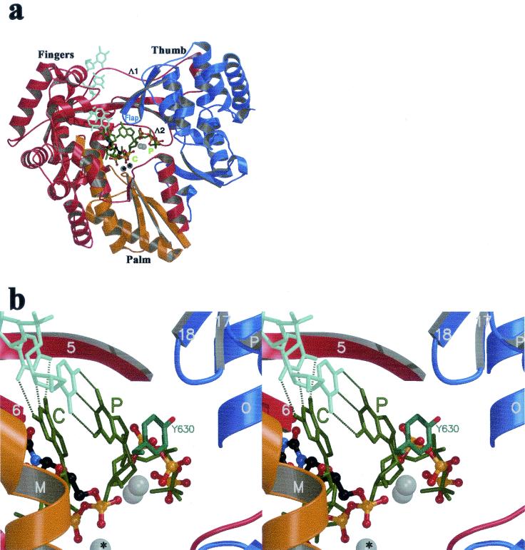
We report here the results of a systematic high-resolution X-ray crystallographic analysis of complexes of the hepatitis C virus (HCV) RNA polymerase with ribonucleoside triphosphates (rNTPs) and divalent metal ions. An unexpected observation revealed by this study is the existence of a specific rGTP binding site in a shallow pocket at the molecular surface of the enzyme, 30 A away from the catalytic site. This previously unidentified rGTP pocket, which lies at the interface between fingers and thumb, may be an allosteric regulatory site and could play a role in allowing alternative interactions between the two domains during a possible conformational change of the enzyme required for efficient initiation. The electron density map at 1.7-A resolution clearly shows the mode of binding of the guanosine moiety to the enzyme. In the catalytic site, density corresponding to the triphosphates of nucleotides bound to the catalytic metals was apparent in each complex with nucleotides. Moreover, a network of triphosphate densities was detected; these densities superpose to the corresponding moieties of the nucleotides observed in the initiation complex reported for the polymerase of bacteriophage phi6, strengthening the proposal that the two enzymes initiate replication de novo by similar mechanisms. No equivalent of the protein stacking platform observed for the priming nucleotide in the phi6 enzyme is present in HCV polymerase, however, again suggesting that a change in conformation of the thumb domain takes place upon template binding to allow for efficient de novo initiation of RNA synthesis.


Figures

FIG. 1.
FIG. 1.
(a and b) The rGTP complex. (a) Overall view, with the finger, palm, and thumb domains colored red, yellow, and blue, respectively. Arrows, β-strands; cylinders, α-helices; thin tubes, connecting loops. The Fo-Fc simulated annealing electron density omit map is displayed at a contour level of 3.5σ as a green net at the surface and C sites. Secondary structure elements next to the surface site (α-helices A, S, and T and loops Λ1 and Λ2) are labeled as described previously (8). This view is from the back of the enzyme, with respect to the views defined in reference . (b) Stereo view of the putative rGTP activator site. The complex has been rotated 90° around a vertical axis with respect to the view shown in panel a, so that Arg32 (at the base of helix A) now lies in the background. The residues involved in contacts with rGTP are displayed in ball-and-stick representation. Two alternative conformations for the sugar and tP of the nucleotide exhibiting about 50% occupancy are represented (with phosphates labeled α, β, and γ and α′, β′, and γ′ denoting the two conformations). Atoms are color coded as follows: carbon, black; nitrogen, blue; oxygen, red; phosphorus, orange; manganese, grey. Broken light blue lines, hydrogen bonds. A thin grey continuous line between one of the guanidinium nitrogens of Arg32 and the N2 position of the guanine base denotes a water-mediated hydrogen bond between the two donors (see text). mc, main chain; w, water molecule. (c) rUTP complex, overall view. The difference electron density with respect to the unliganded enzyme (Fo-Fc simulated annealing omit map contoured at 3σ) is displayed as a green net in the region of the C and P sites. Secondary structure elements relevant to the binding of nucleotides at the C and P sites are labeled.
FIG. 2.
FIG. 2.
The rUTP complex. Secondary structure elements are color coded as in Fig. 1a and labeled as in Fig. 1c. (a) Close-up view of the region around the catalytic site of the enzyme (stereo view). The orientation is as in Fig. 1c, and the representation of atoms is as in Fig. 1b. Amino acids involved in binding the nucleotides are labeled. To the right is the nucleotide bound to the C site, with the uracil base hydrogen bonded to the polypeptide main chain. To the left is the tP moiety of the nucleotide bound at the P site. Hydrogen bonds are represented as in Fig. 1b. Divalent metals (Mn2+) are displayed as gray spheres; *A and *B identify the catalytic ions (see text). mc, main chain. (b) Stereo view displaying the C, P, and I sites (green) in the enzyme's active-site region in the rUTP complex. Shown is a view from the thumb. Labeling is as described in the legend to panel a. Arrow, access route of nucleotides through the NTP tunnel defined previously (8).
FIG. 3.
FIG. 3.
Model for an HCV initiation complex derived from the φ6 polymerase quaternary complex. The images were made after superposition of the equivalent atoms of the HCV and the phage φ6 polymerase (using the equivalent atoms in the fingers and palm defined previously [12]). (a) Stick representation of the template strand (light green) and the two rGTPs (dark green) of the φ6 polymerase initiation complex, along with the rUTP complex (the I site has been removed for clarity). The color code by domains of the protein is the same as that of Fig. 1a; the orientation is the standard front view of the enzyme. The Mn2+ ions are displayed as for Fig. 1 and 2. The C and P sites are labeled in green, with the atoms observed in the rUTP complex displayed as described in Fig. 1a. Flap, β-hairpin 17-18 of the thumb (8). Λ1 and Λ2, loops connecting fingers and thumb at the back of the enzyme. (b) Stereo diagram displaying a close-up view of the P site. Tyr630 of the φ6 polymerase, stacking against the priming base, is displayed in stick representation. Atoms of the rUTP complex are displayed as in Fig. 2a.
FIG. 4.
FIG. 4.
Partial sequence alignment of seven polymerase sequences from HCV with the six major genotypes, with two representatives of HCV with genotype 1 (1a and 1b). Strictly conserved residues are represented as white on a black background. ▾, residues involved in binding the nucleotide at the surface site. A, S, and T, α-helices; arrow (1), β-strand.
FIG. 5.
FIG. 5.
Partial sequence alignment of polymerases of HCV (strain BK), GBV-B (the closest known relative of HCV), flaviviruses (tick-borne encephalitis virus [TBEV] and West Nile virus [WNV]), pestiviruses (BVDV and classical swine fever virus [CSFV]), and bromoviruses (BMV and tomato aspermy virus [TAV]). Secondary structure elements are indicated for the HCV polymerase, with α-helices labeled with capital letters and β-strands labeled with numbers. ▴, residues involved in binding the nucleotide at the priming site. Strictly conserved residues are represented as white on a black background. Residues more than 50% conserved are boxed, with the most commonly found amino acids in bold.

References

    1. Ackermann, M., and R. Padmanabhan. 2001. De novo synthesis of RNA by the dengue virus RNA-dependent RNA polymerase exhibits temperature dependence at the initiation but not elongation phase. J. Biol. Chem. 276:39926-39937. - PubMed
    1. Ago, H., T. Adashi, A. Yoshida, M. Yamamoto, N. Habuka, K. Yatsunami, and M. Miyano. 1999. Crystal structure of the RNA-dependent RNA polymerase of hepatitis C virus. Structure 7:1417-1426. - PubMed
    1. Altschul, S. F., T. L. Madden, A. A. Schaffer, J. Zhang, Z. Zhang, W. Miller, and D. J. Lipman. 1997. Gapped BLAST and PSI-BLAST: a new generation of protein database search programs. Nucleic Acids Res. 25:3389-3402. - PMC - PubMed
    1. Arnold, J. J., and C. E. Cameron. 1999. Poliovirus RNA-dependent RNA polymerase (3Dpol) is sufficient for template switching in vitro. J. Biol. Chem. 274:2706-2716. - PubMed
    1. Arnold, J. J., S. K. Ghosh, and C. E. Cameron. 1999. Poliovirus RNA-dependent RNA polymerase (3D(pol)). Divalent cation modulation of primer, template, and nucleotide selection. J. Biol. Chem. 274:37060-37069. - PubMed

Publication types

MeSH terms

LinkOut - more resources