. 2002 Apr;22(7):2025-36.
doi: 10.1128/MCB.22.7.2025-2036.2002.

Control of cell cycle exit and entry by protein kinase B-regulated forkhead transcription factors

Affiliations

Control of cell cycle exit and entry by protein kinase B-regulated forkhead transcription factors

Geert J P L Kops et al. Mol Cell Biol. 2002 Apr.

Abstract

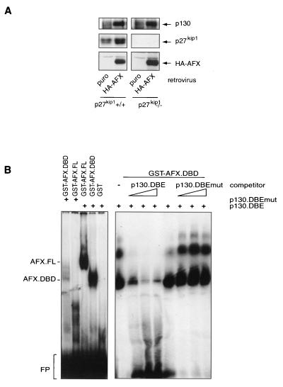
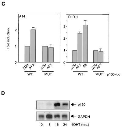
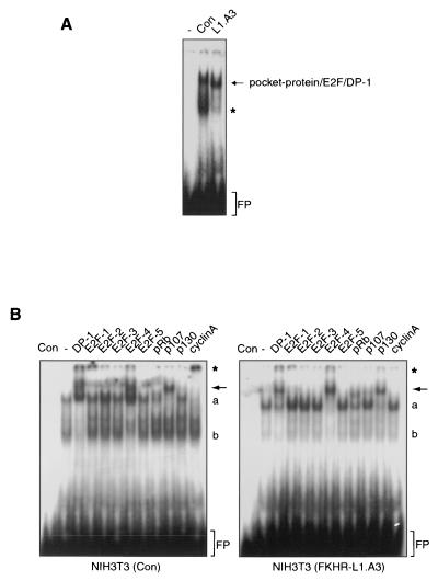
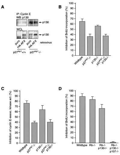
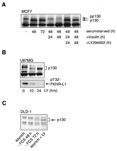
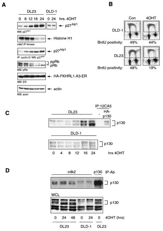
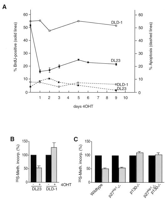
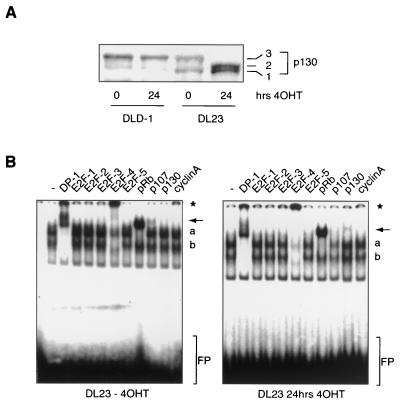
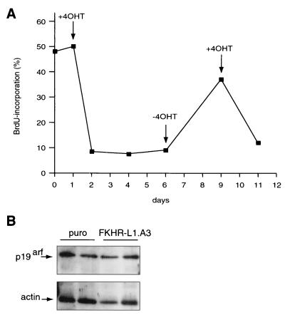
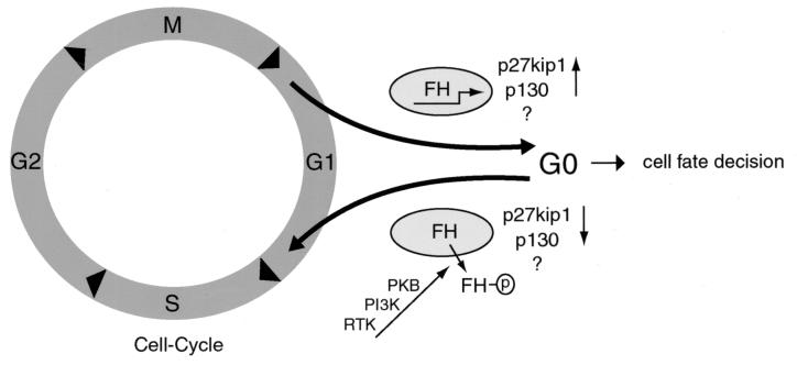
AFX-like Forkhead transcription factors, which are controlled by phosphatidylinositol 3-kinase (PI3K)/protein kinase B (PKB) signaling, are involved in regulating cell cycle progression and cell death. Both cell cycle arrest and induction of apoptosis are mediated in part by transcriptional regulation of p27(kip1). Here we show that the Forkheads AFX (FOXO4) and FKHR-L1 (FOXO3a) also directly control transcription of the retinoblastoma-like p130 protein and cause upregulation of p130 protein expression. Detailed analysis of p130 regulation demonstrates that following Forkhead-induced cell cycle arrest, cells enter G(0) and become quiescent. This is shown by a change in phosphorylation of p130 to G(0)-specific forms and increased p130/E2F-4 complex formation. Most importantly, long-term Forkhead activation causes a sustained but reversible inhibition of proliferation without a marked increase in apoptosis. As for the activity of the Forkheads, we also show that protein levels of p130 are controlled by endogenous PI3K/PKB signaling upon cell cycle reentry. Surprisingly, not only nontransformed cells, but also cancer cells such as human colon carcinoma cells, are forced into quiescence by Forkhead activation. We therefore propose that Forkhead inactivation by PKB signaling in quiescent cells is a crucial step in cell cycle reentry and contributes to the processes of transformation and regeneration.


Figures

FIG. 1.
FIG. 1.
Forkhead transcription factors cause upregulation of the p130 pocket protein. (A) MEFs from wild-type (p27kip1+/+) or p27kip1 knockout (p27kip1−/−) mice were infected with either a control retrovirus or an HA-AFX-containing retrovirus. Total cellular lysates were subsequently analyzed for p130, p27kip1, and HA-AFX expression levels. (B) Purified GST or GST-DBD protein was incubated with a radiolabeled wild-type (p130.DBE) or mutant (p130.DBEmut) probe in the absence or presence of increasing amounts of a nonlabeled oligonucleotide. FL, full length; FP; free probe. (C) A14 or DLD-1 cells were transfected with pGL2-p130intron or pGL2-p130intron.DBEmut, with or without the indicated Forkhead constructs, and luciferase activity was measured. Data are averages from at least three experiments. (D) Two micrograms of RNA was electrophoresed, blotted onto a nylon membrane, and probed for the presence of p130 and GAPDH as a loading control by using radiolabeled probes.
FIG. 1.
FIG. 1.
Forkhead transcription factors cause upregulation of the p130 pocket protein. (A) MEFs from wild-type (p27kip1+/+) or p27kip1 knockout (p27kip1−/−) mice were infected with either a control retrovirus or an HA-AFX-containing retrovirus. Total cellular lysates were subsequently analyzed for p130, p27kip1, and HA-AFX expression levels. (B) Purified GST or GST-DBD protein was incubated with a radiolabeled wild-type (p130.DBE) or mutant (p130.DBEmut) probe in the absence or presence of increasing amounts of a nonlabeled oligonucleotide. FL, full length; FP; free probe. (C) A14 or DLD-1 cells were transfected with pGL2-p130intron or pGL2-p130intron.DBEmut, with or without the indicated Forkhead constructs, and luciferase activity was measured. Data are averages from at least three experiments. (D) Two micrograms of RNA was electrophoresed, blotted onto a nylon membrane, and probed for the presence of p130 and GAPDH as a loading control by using radiolabeled probes.
FIG. 2.
FIG. 2.
Elevated p130 protein levels are indicative of Forkhead-induced cell cycle exit. (A) Total cellular lysates from NIH 3T3 cells that were either left uninfected (−) or infected either with a control virus (Con) or with an FKHR-L1.A3-expressing retrovirus (L1.A3) were analyzed for the presence of E2F/DP-1/pocket protein complexes. Asterisk, free E2F; FP, free probe. (B) NIH 3T3 cells were infected and analyzed as for panel A. Supershifting antibodies are listed above the gel. Arrows indicate the disappearance of p107/E2F-4 complexes in the third lane from the right, and the appearance of p130/E2F-4 complexes in the second lane from the right, due to Forkhead activity. Asterisk, supershifted cyclin A-containing complexes; a, nonsupershifted E2F/pocket protein complexes; b, free E2F; FP, free probe.
FIG. 3.
FIG. 3.
Forkhead-induced upregulation of p130 does not contribute to inhibition of cyclin E-associated kinase activity. (A) MEFs infected as for Fig. 1A were lysed, and the amount of p130 in cyclin E-containing complexes was analyzed by Western blotting (WB) of cyclin E immunoprecipitates (IP). WCL, whole-cell lysate. (B and C) Primary wild-type MEFs and MEFs from p27kip1, p130, or p27kip1 p130 knockout mice were infected with a control or an FKHR-L1.A3-encoding retrovirus. At 24 h postinfection, proliferation was measured by BrdU incorporation (B) and cyclin E-associated kinase activity (act.) (C). Graphs show relative inhibition of proliferation by FKHR-L1.A3, as measured by these two assays, compared to proliferation with the control. Data are averages from three independent experiments. (D) Primary wild-type MEFs and MEFs from pRb, pRb p130, and pRb p130 p107 knockout mice were treated with 10 nM LY249002 for 16 h, and proliferation was assayed by BrdU incorporation.
FIG. 4.
FIG. 4.
Forkhead-induced cell cycle exit is mediated by PI3K signaling. (A) MCF7 cells were serum starved for 48 or 72 h and subsequently treated with insulin for 24 or 48 h, with or without a 30-min preincubation with LY294002. p130 protein levels were then analyzed by Western blotting. pp130, hyperphosphorylated p130. (B) U87MG cells were treated with 10 μM LY294002 (LY) for 0, 10, or 24 h. Total cellular lysates were immunoblotted for the presence of p130 protein and T32-phosphorylated FKHR-L1. (C) p130 protein levels of asynchronous (asynch) DLD-1 cells were compared to those of DLD-1 cells that were either serum starved (− FCS) for 48 or 72 h or treated with LY294002 for 16 h.
FIG. 5.
FIG. 5.
Conditional activation of FKHR-L1 causes growth arrest and increased p130 protein levels in human colon carcinoma cells. (A) DL23 and DLD-1 cells were left untreated (0 h) or treated with 500 nM 4OHT for 8, 12, 16, or 24 h. Total cellular lysates were electrophoresed and blotted for the presence of p27kip1, pRb, HA-FKHR-L1.A3-ER,or actin. Cell lysates were analyzed for cdk2 kinase activity by cdk2 immunoprecipitation (IP) and in vitro kinase reaction using histone H1 as a substrate. The cdk2 kinase reaction products were blotted, autoradiographed, and probed for the presence of p27kip1. WB, Western blot. (B) BrdU incorporation of DL23 and DLD-1 cells left untreated (Con) or treated with 500 nM 4OHT for 16 h. (C) Total cellular lysates from DLD-1 and DL23 cells treated without (0 h) or with 500 nM 4OHT for 4, 8, 12, 16, or 24 h were analyzed by Western blotting for the presence of the p130 retinoblastoma-like protein. Anti-HA immunoprecipitations of A14 cells transiently transfected with empty vector or pCMV-HA-p130 served as controls. (D) DL23 and DLD-1 cells, left untreated (0 h) or treated with 4OHT for 24 or 48 h, were lysed, and cdk2 was immunoprecipitated. The cdk2 immunoprecipitates were subsequently analyzed for the presence of p130 by Western blotting. WCL, whole-cell lysate; IP-Ab, immunoprecipitating antibody.
FIG. 6.
FIG. 6.
Long-term activation of FKHR-L1 causes continued arrest.(A) DLD-1 and DL23 cells were treated with 500 nM 4OHT for 9 days. Cells were seeded to approximately 5% confluency. Cells were given fresh 4OHT-containing medium every day and were split whenever cell density reached 70 to 80%. Typically, during the whole experiment, only the DLD-1 control cells were split and seeded again to 5% confluency (usually at day 5). Samples were taken at various time points to measure proliferation by BrdU incorporation (solid lines) and cell death by FACS analysis of sub-G1 population (dashed lines). (B) DLD-1 and DL23 cells were left untreated (−) or treated with 500 nM 4OHT for 48 h (+) and pulse-labeled with [35S]methionine to measure protein synthesis. (C) Primary wild-type MEFs and MEFs from p27kip1, p130, or p27kip1 p130 knockout mice were infected with control virus (pBabe-puro) (solid bars) or FKHR-L1.A3-encoding retrovirus (shaded bars) and pulse-labeled with [35S]methionine at 24 h postinfection to measure protein synthesis.
FIG. 7.
FIG. 7.
Specific activation of FKHR-L1 causes human carcinoma cells to exit the cell cycle. (A) Total cellular lysates from DLD-1 and DL23 cells that were left untreated (0 h) or treated with 500 nM 4OHT for 24 h were electrophoresed on a 6% polyacrylamide gel and immunoblotted for the presence of p130. The three phosphorylated forms of the p130 protein are indicated as 1, 2, and 3. (B) Total cellular lysates from DL23 cells left untreated (left) or treated with 500 nM 4OHT for 48 h (right) were analyzed for the presence of E2F/DP-1/pocket protein complexes. Supershifting antibodies are listed above the gel. Arrows indicate the disappearance of p107/E2F-4 complexes in the third lane from the right and the appearance of p130/E2F-4 complexes in the second lane from the right upon 4OHT treatment. Asterisk, supershifted E2F-4- or cyclin A-containing complexes; a, nonsupershifted E2F/pocket protein complexes; b, free E2F; FP, free probe.
FIG. 8.
FIG. 8.
Forkhead-induced G0 is reversible. (A) DL23 cells were seeded to approximately 5% confluency. Confluency was monitored during the experiment, as described for Fig. 6A, but none of the dishes at any of the indicated time points required splitting. Cells were given fresh medium without 4OHT (−4OHT) or with 500 nM 4OHT (+4OHT) every day. When 4OHT was removed (at day 6), cells were washed twice with medium without 4OHT before the fresh medium was added. Similarly, when cells were again given 4OHT (at day 9), they were washed twice with medium containing 4OHT before the fresh medium was added. Samples were taken at days 0, 1, 2, 3, 4, 6, 9, and 11, and BrdU incorporation was measured. (B) Samples of primary wild-type MEFs from two independent infections with either a control virus (puro) or an FKHR-L1.A3-encoding retrovirus were analyzed for p19arf expression by immunoblotting.
FIG. 9.
FIG. 9.
Model for cell cycle regulation by the PI3K/PKB/Forkhead pathway. When cells do not receive growth factors in early G1, PKB is not activated and Forkheads (FH) remain active. This results in upregulation of the genes for p27kip1 and p130 along with yet unidentified target genes, leading to cell cycle exit in nonhematopoietic cells and, with the additional regulation of the Bim and FasL gene products, to apoptosis in hematopoietic cells. Subsequent activation of PKB through receptor tyrosine kinases (RTK) leads to phosphorylation of FH (indicated by the circled p) and inactivation of Forkheads by nuclear exclusion and therefore to cell cycle reentry via downregulation of the Forkhead cell cycle-regulatory target genes. See the text for further details.

References

    1. Albrecht, J. H., R. Y. Poon, C. L. Ahonen, B. M. Rieland, C. Deng, and G. S. Crary. 1998. Involvement of p21 and p27 in the regulation of CDK activity and cell cycle progression in the regenerating liver. Oncogene 16:2141-2150. - PubMed
    1. Baldi, A., V. Esposito, A. De Luca, C. M. Howard, G. Mazzarella, F. Baldi, M. Caputi, and A. Giordano. 1996. Differential expression of the retinoblastoma gene family members pRb/p105, p107, and pRb2/p130 in lung cancer. Clin. Cancer Res. 2:1239-1245. - PubMed
    1. Bos, J. L. 1998. All in the family? New insights and questions regarding interconnectivity of Ras, Rap1 and Ral. EMBO J. 17:6776-6782. - PMC - PubMed
    1. Bos, J. L. 1995. p21ras: an oncoprotein functioning in growth factor-induced signal transduction. Eur. J. Cancer 31:1051-1054. - PubMed
    1. Bouzahzah, B., M. Fu, A. Iavarone, V. M. Factor, S. S. Thorgeirsson, and R. G. Pestell. 2000. Transforming growth factor-β1 recruits histone deacetylase 1 to a p130 repressor complex in transgenic mice in vivo. Cancer Res. 60:4531-4537. - PubMed

Publication types

MeSH terms

Substances