. 2002 Apr;22(7):2047-56.
doi: 10.1128/MCB.22.7.2047-2056.2002.

Ime2, a meiosis-specific kinase in yeast, is required for destabilization of its transcriptional activator, Ime1

Affiliations

Ime2, a meiosis-specific kinase in yeast, is required for destabilization of its transcriptional activator, Ime1

Noga Guttmann-Raviv et al. Mol Cell Biol. 2002 Apr.

Abstract

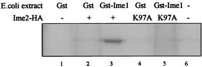
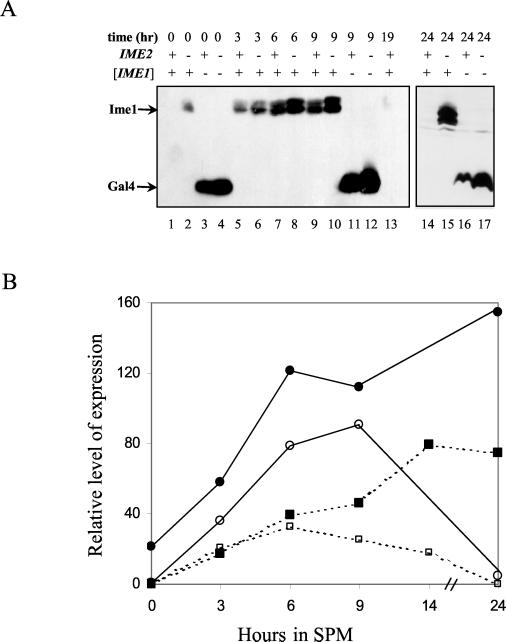
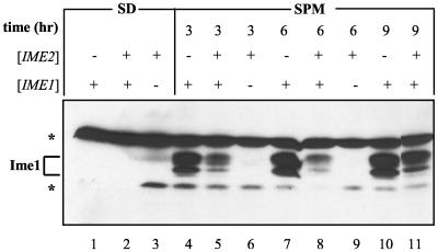
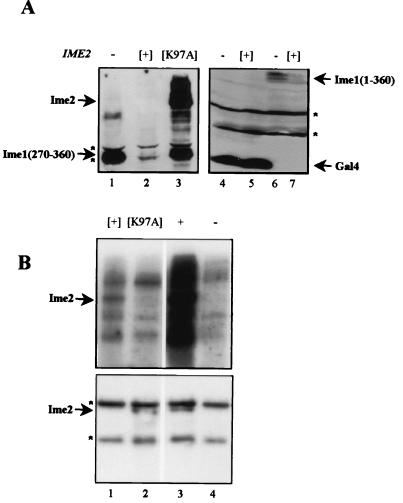
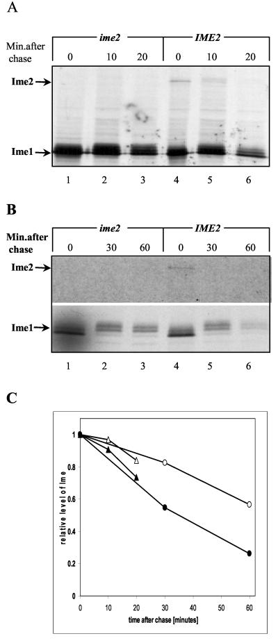
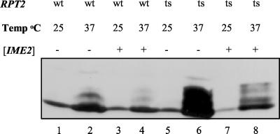
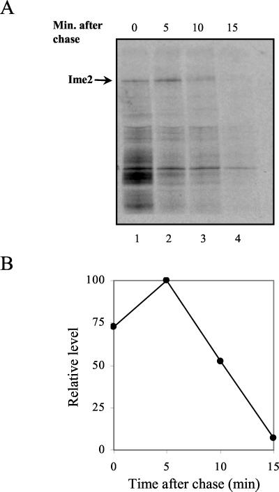
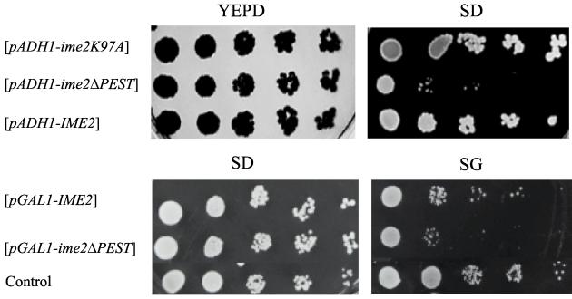
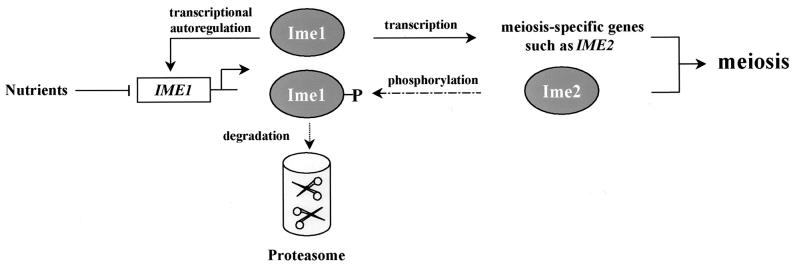
In the budding yeast Saccharomyces cerevisiae, entry into meiosis and its successful completion depend on two positive regulators, Ime1 and Ime2. Ime1 is a transcriptional activator that is required for transcription of IME2, a serine/threonine protein kinase. We show that in vivo Ime2 associates with Ime1, that in vitro Ime2 phosphorylates Ime1, and that in living cells the stability of Ime1 depends on Ime2. Diploid cells with IME2 deleted show an increase in the level of Ime1, whereas haploid cells overexpressing IME2 show a decrease in the stability of Ime1. Furthermore, the level of Ime1 depends on the kinase activity of Ime2. Using a mutation in one of the ATPase subunits of the proteasome, RPT2, we demonstrate that Ime1, amino acids 270 to 360, is degraded by the 26S proteasome. We also show that Ime2 itself is an extremely unstable protein whose expression in vegetative cultures is toxic. We propose that a negative-feedback loop ensures that the activity of Ime1 will be restricted to a narrow window.


Figures

FIG. 1.
FIG. 1.
Ime2 phosphorylates Ime1 in vitro. Yeast cells (strain Y742) carrying either pGAL1-3HA-IME2(1-645) (YEp1985) (lanes 2 and 3), pGAL1-3HA-ime2K97A(1-645) (YEp2264) (lanes 4 and 5), or pGAL1-3HA (YEp1906) (lanes 1 and 6) grown in SR medium to 107 cells/ml were transferred to SG medium for 16 h. Proteins were extracted and immunoprecipitated with antibodies directed against 12CA5. GST-Ime1(270-360) (lanes 2 and 4) and Gst1 (lanes 1 and 3) isolated on glutathione-Sepharose beads were added to the immunoprecipitated extracts, subjected to a kinase reaction, and separated on an SDS-10% PAGE gel.
FIG. 2.
FIG. 2.
Ime2 modulates the level of Ime1 in diploid cells. Proteins were extracted from meiotic cultures at 0 (lanes 1 to 4), 3 (lanes 5 to 6), 6 (lanes 7 to 8), 9 (lanes 9 to 12), 19 (lane 13), and 24 (lanes 14 to 17) h of incubation in SPM. Strains used were MATa/MATα IME2/IME2 (Y422) (lanes 1, 3, 5, 7, 9, 11, 13, 14, and 16) and MATa/MATα ime2Δ/ime2Δ (Y1039) (lanes 2, 4, 6, 8, 10, 12, 15, and 17). (A) Cells carried the following genes on a 2μm plasmid: pIME1-gal4(1-147)-ime1(270-360) (P1931) (lanes 1, 2, 5 to 10, and 13 to 15), and pIME1-gal4(1-147) (P1952) (lanes 3, 4, 11, 12, 16, and 17). Immunoblot analysis was performed by using antibodies directed against Gal4(bd). Brackets indicate that the gene was present on a multicopy plasmid. (B) The relative levels of Ime1 (solid lines and circles) were quantitated from the results shown in panel A, and those of Ime1-LacZ (dashed lines and squares) were calculated from β-galactosidase assays on three transformants carrying pAS128. Solid symbols, strain Y422; open symbols, strain Y1039.
FIG. 3.
FIG. 3.
Ime2 modulates the level of Ime1 in haploid cells. Proteins were extracted either from cells grown in SD medium to 107 cells/ml (lanes 1 to 3) or from meiotic cultures at 3 (lanes 4 to 6), 6 (lanes 7 to 9), or 9 (lanes 10 and 11) h in SPM. A MATa ime2Δ strain (Y1071) carrying the following genes on a 2μm plasmid was used: pIME1-gal4(1-147)-HA-IME2 (P1968) (lanes 3, 6, and 9), pIME1-gal4(1-147)-ime1(270-360) (P1931) (lanes 1, 4, 7, and 10), or both P1968 and P1931 (lanes 2, 5, 8, and 11). Immunoblot analysis was performed using antibodies directed against Gal4(bd). Brackets indicate that the gene was present on a multicopy plasmid. Asterisks, nonspecific bands.
FIG. 4.
FIG. 4.
The stability of Ime1 depends on the kinase activity of Ime2. (A) Western blot analysis. Proteins were extracted from cells grown in SD medium to 107 cells/ml and loaded onto an SDS-PAGE gel, and immunoblot analysis was performed using antibodies directed against Gal4(bd). A MATa ime2Δ strain (Y1071) carrying either pADH1-gal4(1-147)-ime1(270-360) (YEp1969) (lanes 1 to 3), pADH1-gal4(1-147) [pGBDU-C(1)] (lanes 4 to 5), or pADH1-gal4(1-147)-ime1(1-360) (YEp1202) (lanes 6 to 7) was used. This strain also carried any one of the following plasmids: YEplac112 (14) (lanes 1, 4, and 6) (control), YEp1916 (lanes 2, 5, and 7) (pADH1-gal4(1-147)-HA-IME2), and P2225 (lane 3) (pADH1-gal4(1-147)-ime2K97A. (B) Cells grown in SR medium to 107/ml were transferred to SG medium for 16 h. Proteins were extracted, immunoprecipitated with antibodies directed against 12CA5, subjected to a kinase reaction, and separated on an SDS-10% PAGE gel. (Upper panel) kinase assay; (lower panel) IP-Western. Strains used were MATa LEU2::pGAL1-3HA-IME2(1-645) (Y1255) (lane 3) and its isogenic MATa strain (Y742) carrying pGAL1-3HA-IME2(1-645) (YEp1985) (lane 1), pGAL1-3HA-ime2K97A (YEp2264) (lane 2), and pGAL1-3HA (YEp1906) (lane 4). Brackets indicate that the gene was present on a multicopy plasmid. Asterisks, nonspecific bands.
FIG. 5.
FIG. 5.
Ime2 regulates the half-life of Ime1. Shown are results of a pulse (7 min)-chase analysis of Ime1(id) and Ime2 tagged with Gal4(bd). (A) Proteins were extracted from cells grown to a density of 107/ml in SD medium. A MATa ime2Δ strain (Y1071) carrying pADH1-gal4(1-147)-ime1(270-360) on a 2μm plasmid (P1969) (lanes 1 to 6) or pADH1-gal4(1-147)-IME2 on a 2μm plasmid (P1916) (lanes 4 to 6) was used. (B) Proteins were extracted from cells grown in SD medium to stationary phase, washed in water, and incubated in SPM at a titer of 107 cells/ml for 4 h. A MATa ime2Δ strain (Y1071) carrying pIME1-gal4(1-147)-ime1(270-360) (P1931) (lanes 1 to 6) or pIME1-gal4(1-147)-IME2 (P1968) (lanes 4 to 6) was used. The film exposure was 15 times longer for the meiotic culture than for the mitotic one. This phenomenon is typical for meiotic cultures that become nonpermissive for the incorporation of compounds from the media (23). (C) The intensities of the bands corresponding to Gal4(bd)-Ime1(id) were quantitated by PhosphorImager analysis and are expressed as percentages of those present during initiation of the chase. Triangles, vegetative cultures; circles, meiotic cultures. Open symbols, cells with deletions of IME2; solid symbols, cells overexpressing Ime2. Brackets indicate that the gene was present on a multicopy plasmid.
FIG. 6.
FIG. 6.
Ime1 is degraded by the proteasome. Proteins were extracted from cells that were grown in SD medium at 25°C (lanes 1, 3, 5, and 7) and incubated for 6 h at 37°C (lanes 2, 4, 6, and 8). Strains used were Y1248 (lanes 1 to 4) and its isogenic rpt2RF strain (Y1249) (lanes 5 to 8) carrying YEp1974 [pADH1-gal4(1-147)-ime1(270-360)] and YEpLac112 (14) (control) (lanes 1, 2, 5, and 6) or YEp1974 and YEp1916 [pADH1-gal4(1-147)-HA-IME2] (lanes 3, 4, 7, and 8). Immunoblot analysis was performed using antibodies directed against Gal4(bd). Brackets indicate that the gene was present on a multicopy plasmid.
FIG. 7.
FIG. 7.
IME2 encodes an unstable protein. Pulse (4 min)-chase analysis of Ime2 tagged with Gal4(bd). (A) Proteins were extracted from meiotic cultures at 4 h in SPM. A MATa strain (sub62) (38) carrying pIME1-IME2 on a 2μm plasmid (YEp1968) was used. (B) Quantitative analysis of the level of Ime2.
FIG. 8.
FIG. 8.
Overexpression of Ime2 in vegetative cultures is toxic. MATa strains (Y742) carrying various IME2 plasmids grown on SD plates were plated on YEPD (rich glucose medium), SD (minimal glucose medium), and SG (minimal galactose medium) plates following serial 10-fold dilutions. Plasmids used were YEp1916 (pADH1-IME2), YEp2126 (pADH1-ime2ΔPEST), YEp2225 (pADH1-ime2K97A), YEp1985 (pGAL1-IME2), and YEp2142 (pGAL1-ime2ΔPEST). As a control, Y742 carried the vector plasmid, YEpLac112 (14). Brackets indicate that the gene was present on a multicopy plasmid.
FIG. 9.
FIG. 9.
A feedback loop controlling meiosis. Shown is a schematic illustration of the transcriptional cascade and feedback loops that control entry into meiosis.

References

    1. Bowdish, K. S., H. E. Yuan, and A. P. Mitchell. 1994. Analysis of RIM11, a yeast protein kinase that phosphorylates the meiotic activator IME1. Mol. Cell. Biol. 14:7909-7919. - PMC - PubMed
    1. Bradford, M. M. 1976. A rapid and sensitive method for the quantitation of microgram quantities of protein utilizing the principle of protein-dye binding. Anal. Biochem. 72:248-254. - PubMed
    1. Chien, C. T., P. L. Bartel, R. Sternglanz, and S. Fields. 1991. The two-hybrid system: a method to identify and clone genes for proteins that interact with a protein of interest. Proc. Natl. Acad. Sci. USA 88:9578-9582. - PMC - PubMed
    1. Chu, S., J. DeRisi, M. Eisen, J. Mulholland, D. Botstein, P. O. Brown, and I. Herskowitz. 1998. The transcriptional program of sporulation in budding yeast. Science 282:699-705. (Erratum, 282:1421.) - PubMed
    1. Ciechanover, A. 1998. The ubiquitin-proteasome pathway: on protein death and cell life. EMBO J. 17:7151-7160. - PMC - PubMed

Publication types

MeSH terms

LinkOut - more resources