. 2002 Apr;22(7):2099-110.
doi: 10.1128/MCB.22.7.2099-2110.2002.

Convergence of multiple signaling cascades at glycogen synthase kinase 3: Edg receptor-mediated phosphorylation and inactivation by lysophosphatidic acid through a protein kinase C-dependent intracellular pathway

Affiliations

Convergence of multiple signaling cascades at glycogen synthase kinase 3: Edg receptor-mediated phosphorylation and inactivation by lysophosphatidic acid through a protein kinase C-dependent intracellular pathway

Xianjun Fang et al. Mol Cell Biol. 2002 Apr.

Abstract

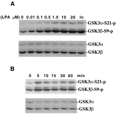
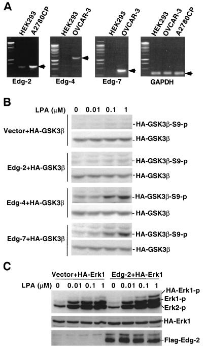
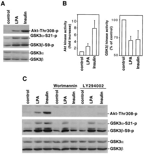
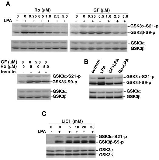
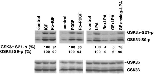
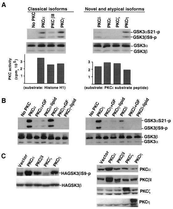
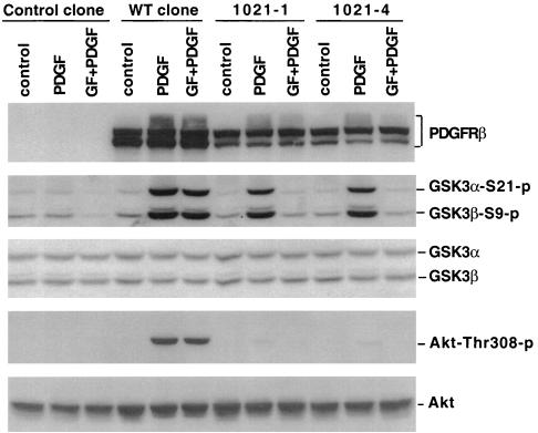
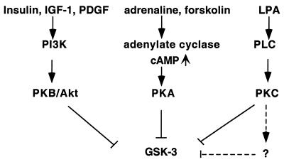
Lysophosphatidic acid (LPA) is a natural phospholipid with multiple biological functions. We show here that LPA induces phosphorylation and inactivation of glycogen synthase kinase 3 (GSK-3), a multifunctional serine/threonine kinase. The effect of LPA can be reconstituted by expression of Edg-4 or Edg-7 in cells lacking LPA responses. Compared to insulin, LPA stimulates only modest phosphatidylinositol 3-kinase (PI3K)-dependent activation of protein kinase B (PKB/Akt) that does not correlate with the magnitude of GSK-3 phosphorylation induced by LPA. PI3K inhibitors block insulin- but not LPA-induced GSK-3 phosphorylation. In contrast, the effect of LPA, but not that of insulin or platelet-derived growth factor (PDGF), is sensitive to protein kinase C (PKC) inhibitors. Downregulation of endogenous PKC activity selectively reduces LPA-mediated GSK-3 phosphorylation. Furthermore, several PKC isotypes phosphorylate GSK-3 in vitro and in vivo. To confirm a specific role for PKC in regulation of GSK-3, we further studied signaling properties of PDGF receptor beta subunit (PDGFRbeta) in HEK293 cells lacking endogenous PDGF receptors. In clones expressing a PDGFRbeta mutant wherein the residues that couple to PI3K and other signaling functions are mutated with the link to phospholipase Cgamma (PLCgamma) left intact, PDGF is fully capable of stimulating GSK-3 phosphorylation. The process is sensitive to PKC inhibitors in contrast to the response through the wild-type PDGFRbeta. Therefore, growth factors, such as PDGF, which control GSK-3 mainly through the PI3K-PKB/Akt module, possess the ability to regulate GSK-3 through an alternative, redundant PLCgamma-PKC pathway. LPA and potentially other natural ligands primarily utilize a PKC-dependent pathway to modulate GSK-3.


Figures

FIG. 1.
FIG. 1.
LPA induces phosphorylation of GSK-3α at serine 21 (S21) and GSK-3β at serine 9 (S9) in a dosage- and time-dependent manner. Serum-starved Swiss 3T3 cells were stimulated with LPA. After stimulation, cell lysates were prepared and analyzed for GSK-3 phosphorylation by Western blotting with a GSK-3 phospho-specific antibody as detailed in Materials and Methods. Reprobing with a phosphorylation-independent antibody against total GSK-3α and GSK-3β was included to show equal loading among samples. (A) Cells were stimulated for 5 min with indicated concentrations of LPA or with insulin (In, 0.1 μg/ml). (B) Cells were incubated with 10 μM LPA for different periods of time (minutes) as indicated. Experiments were performed at least three times with consistent results.
FIG. 2.
FIG. 2.
LPA-induced GSK-3 phosphorylation is mediated by Edg-4 or Edg-7 but not Edg-2. (A) Total cellular RNA was isolated from HEK293, A2780CP, and OVCAR-3 cells. Expression of Edg-2, Edg-4, Edg-7, and GAPDH mRNA was analyzed by RT-PCR as described in Materials and Methods. PCR products consistent with expected sizes are indicated by arrows. (B) HEK293 cells were cotransfected with HA-GSK-3β, along with an empty vector or an Edg expression vector. Two days after transfection, cells were starved for 8 h and stimulated with LPA at indicated concentrations for 5 min. Cell lysates were prepared and analyzed for GSK-3β phosphorylation by immunoblotting with a GSK-3β phospho-specific antibody that reacts only with GSK-3β phosphorylated at serine 9. Expression of transfected HA-GSK-3β was verified by immunoblotting with an anti-HA monoclonal antibody. (C) HEK293 cells were cotransfected with HA-Erk1, along with an empty vector or Edg-2 expression vector. Cells were treated as in panel B, and lysates were analyzed for Erk phosphorylation by immunoblotting with an Erk phospho-specific antibody. Expression of Flag-Edg-2 in transfected cells was confirmed by immunoblotting with anti-Flag-M2 antibody. Similar results were obtained from three independent experiments.
FIG. 3.
FIG. 3.
LPA-induced GSK-3 phosphorylation does not correlate with activation of the PI3K-PKB/Akt cascade. (A) Serum-starved Swiss 3T3 cells were stimulated with LPA (10 μM, 5 min) or insulin (0.1 μg/ml, 5 min). Cell lysates were prepared and analyzed for phosphorylation of PKB/Akt (Thr-308) and GSK-3 by immunoblotting with PKB/Akt or GSK-3 phospho-specific antibodies. Reprobing with an antibody against total GSK-3α and GSK-3β was performed to show equal loading among samples. Shown is a representative experiment of four independent assays. (B) PKB/Akt and GSK-3β kinase activity in LPA- and insulin-stimulated cells was measured by in vitro kinase assays as described in Materials and Methods. PKB/Akt activity was quantified by densitometry as reported previously (21) and presented as fold increases over unstimulated control cells. GSK-3β activity was presented as percentages of the activity of unstimulated control cells. The results for both PKB/Akt and GSK-3β are means ± the standard deviation of three independent experiments. (C) Swiss 3T3 cells were stimulated with LPA (10 μM) or insulin (0.1 μg/ml) for 5 min in the presence of the PI3K inhibitors, wortmannin (0.1 μM), or LY294002 (10 μM) or vehicle. Wortmannin, LY294002, or vehicle was added to culture 45 to 60 min before stimulation. Cell lysates were analyzed for PKB/Akt and GSK-3 phosphorylation as in panel A.
FIG. 4.
FIG. 4.
LPA-induced GSK-3 phosphorylation is sensitive to PKC inhibitors. (A) Serum-starved Swiss 3T3 cells were stimulated with LPA (10 μM, 5 min) in the absence or presence of indicated concentrations of the PKC inhibitor Ro31-8220 (Ro) or GF109203X (GF). In the bottom panel, Swiss 3T3 cells were stimulated with insulin (0.1 μg/ml, 5 min) without or with Ro31-8220 (Ro, 5 μM) or GF109203X (GF, 5 μM). Ro31-8220 or GF109203X was added to culture where indicated 45 to 60 min before stimulation. Cell lysates were prepared and analyzed for GSK-3α and GSK-3β phosphorylation by immunoblotting with a GSK-3 phospho-specific antibody as described in Fig. 1. Shown are representative experiments of three independent assays. (B) Swiss 3T3 cells were stimulated with LPA (10 μM) for 1 h with or without GF109203 (GF, 2.5 μM) or Ro31-8220 (2.0 μM). Cell lysates were analyzed for GSK-3 phosphorylation as in panel A. (C) Swiss 3T3 cells were stimulated with LPA (10 μM, 5 min) in the presence of lithium chloride (LiCl) at the indicated concentrations. LiCl was added to medium 75 min before LPA stimulation. Cell lysates were prepared and analyzed as in panel A.
FIG. 5.
FIG. 5.
PKC inhibitors do not block GSK-3 phosphorylation induced by peptide growth factors. Serum-starved Swiss 3T3 cells were stimulated for 10 min with IGF-1 (50 ng/ml), PDGF (50 ng/ml), or LPA (10 μM) in the presence of Ro31-8220 (Ro, 2.25 μM), GF109203X (GF, 2.0 μM), the GF109203X inactive analog bisindolylmaleimide V (GF analog, 2.0 μM), or vehicle. These compounds were added to culture where indicated 45 to 60 min before stimulation with IGF-1, PDGF, or LPA. Cell lysates were prepared and analyzed for GSK-3α and GSK-3β phosphorylation by immunoblotting as described in Fig. 1. The bands representing phosphorylated α or GSK-3β were quantified by densitometry. The numbers represent the percent relative intensities, with the values of unstimulated cells (control) defined as background and the values of cells stimulated in the absence of PKC inhibitors minus background defined as 100%. Similar results were obtained from three independent experiments.
FIG. 6.
FIG. 6.
TPA induces GSK-3 phosphorylation, and chronic treatment with TPA compromises LPA-mediated GSK-3 phosphorylation. (A) Serum-starved Swiss 3T3 cells were stimulated with TPA for 10 min at the indicated concentrations. In the right panel, cells were stimulated with TPA (0.1 μM, 10 min) in the presence of Ro31-8220 (Ro, 4.5 μM), GF109203X (GF, 2 μM), or vehicle. Ro31-8220, GF109203X, or vehicle was added to culture 45 to 60 min before stimulation with TPA. (B) Swiss 3T3 cells were preincubated with 0.2 μM TPA or vehicle in serum-free medium for more than 24 h before stimulation with LPA (10 μM, 5 min), IGF-1 (50 ng/ml, 5 min), or TPA (0.1 μM, 10 min). For both panels A and B, cell lysates were prepared and analyzed for GSK-3α and GSK-3β phosphorylation by immunoblotting as in Fig. 1. Similar results were obtained in three independent experiments.
FIG. 7.
FIG. 7.
Specific PKC isotypes can phosphorylate GSK-3α at serine 21 and GSK-3β at serine 9 in vitro and in vivo. (A) Different isoforms of PKC were incubated with purified GSK-3 in an in vitro kinase assay as described in Materials and Methods. Reaction mixes were diluted in SDS sample buffer and analyzed for phosphorylation of GSK-3α at serine 21 and of GSK-3β at serine 9 by immunoblotting with a GSK-3 phospho-specific antibody as described in Fig. 1. Reprobing with a phosphorylation-independent antibody against total GSK-3α and GSK-3β (substrates) was included to show equal loading among samples. The bottom panel shows activity of the PKC isotypes toward standard PKC substrates (histone H1 for classical forms and PKCɛ substrate peptide for novel and atypical forms). (B) PKCα, PKCγ, PKCη, and PKCδ were incubated with GSK-3 in the presence of the PKC inhibitor GF109203X (+GF, 0.15 μM for PKCα and PKCγ, 0.6 μM for PKCη and PKCδ) or without lipid activators (−lipid) as indicated. The reaction mixes were analyzed as in panel A. (C) HEK293 cells were cotransfected with HA-GSK-3β, along with an empty vector or a constitutively active PKC construct as indicated. Two days after transfection, cells were starved for 12 h, and lysates were prepared and analyzed for HA-GSK-3β phosphorylation levels with a GSK-3β phospho-specific antibody. Expression of transfected HA-GSK-3β was detected by immunoblotting with an anti-HA monoclonal antibody. Expression of PKC proteins was confirmed by immunoblotting with PKC isotype-specific antibodies. The anti-PKCβ1 antibody cross-reacts with PKCα, whereas the other anti-PKC antibodies did not exhibit cross-reactivity. Each of these experiments was performed at least twice with consistent results.
FIG. 8.
FIG. 8.
The PLCγ pathway is sufficient for PDGF-induced GSK-3 phosphorylation. HEK293 cells were transfected with an empty backbone vector, WT PDGFRβ, or mutant PDGFRβ (Y-1021). Stable clones carrying empty vector (control clone) or expressing WT PDGFRβ (WT clone) or mutant PDGFRβ (Y-1021-1 and Y-1021-4) were established. After starvation in serum-free medium for more than 12 h, these clones were stimulated with PDGF B/B (75 ng/ml, 10 min) in the presence of GF109203X (GF, 2.5 μM) or vehicle. Cells were preincubated with GF109203X or vehicle for 45 to 60 min before stimulation with PDGF B/B. Cell lysates were analyzed for phosphorylation of GSK-3 and PKB/Akt (threonine 308) by immunoblotting as described in Materials and Methods. Reprobing with phosphorylation-independent antibodies against total GSK-3α and GSK-3β or total PKB/Akt was included to show equal loading among samples. The expression of PDGFRβ in WT and Y-1021 clones, but not in the control clone, was verified by immunoblotting with an anti-PDGFRβ antibody. Similar results were obtained in three independent experiments
FIG. 9.
FIG. 9.
Convergence of multiple intracellular signaling pathways at GSK-3.

Similar articles

Cited by

References

    1. Adams, J. C., and W. J. Gullick. 1989. Differences in phorbol-ester-induced downregulation of protein kinase C between cell lines. Biochem. J. 257:905-911. - PMC - PubMed
    1. Bellacosa, A., J. R. Testa, S. P. Staal, and P. N. Tsichlis. 1991. A retroviral oncogene, akt, encoding a serine-threonine kinase containing an SH2-like region. Science 254:271-274. - PubMed
    1. Bonni, A., A. Brunet, A. E. West, S. R. Datta, M. A. Takasu, and M. E. Greenberg. 1999. Cell survival promoted by the Ras-MAPK signaling pathway by transcription-dependent and independent mechanisms. Science 286:1358-1362. - PubMed
    1. Cai, H., U. Smola, V. Wixler, I. Eisenmann-Tappe, M. T. Diaz-Meco, J. Moscat, U. Rapp, and G. M. Cooper. 1997. Role of diacylglycerol-regulated protein kinase C isotypes in growth factor activation of the Raf-1 protein kinase. Mol. Cell. Biol. 17:732-741. - PMC - PubMed
    1. Chen, E. Y., N. M. Mazure, J. A. Cooper, and A. J. Giaccia. 2001. Hypoxia activates a platelet-derived growth factor receptor/phosphatidylinositol 3-kinase/Akt pathway that results in glycogen synthase kinase-3 inactivation. Cancer Res. 61:2429-2433. - PubMed

Publication types

MeSH terms

LinkOut - more resources