. 2002 Apr;22(7):2170-81.
doi: 10.1128/MCB.22.7.2170-2181.2002.

Functional divergence between histone deacetylases in fission yeast by distinct cellular localization and in vivo specificity

Affiliations

Functional divergence between histone deacetylases in fission yeast by distinct cellular localization and in vivo specificity

Pernilla Bjerling et al. Mol Cell Biol. 2002 Apr.

Erratum in

  • Mol Cell Biol 2002 Jul;22(14):5257-8

Abstract

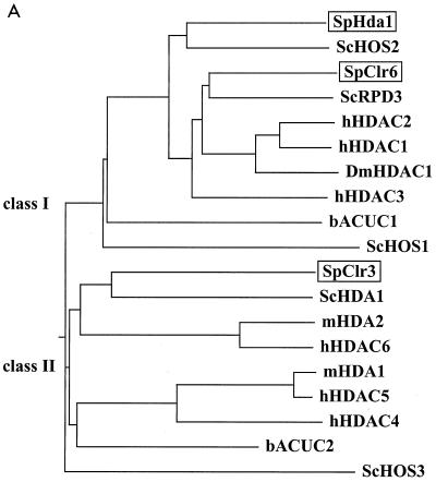
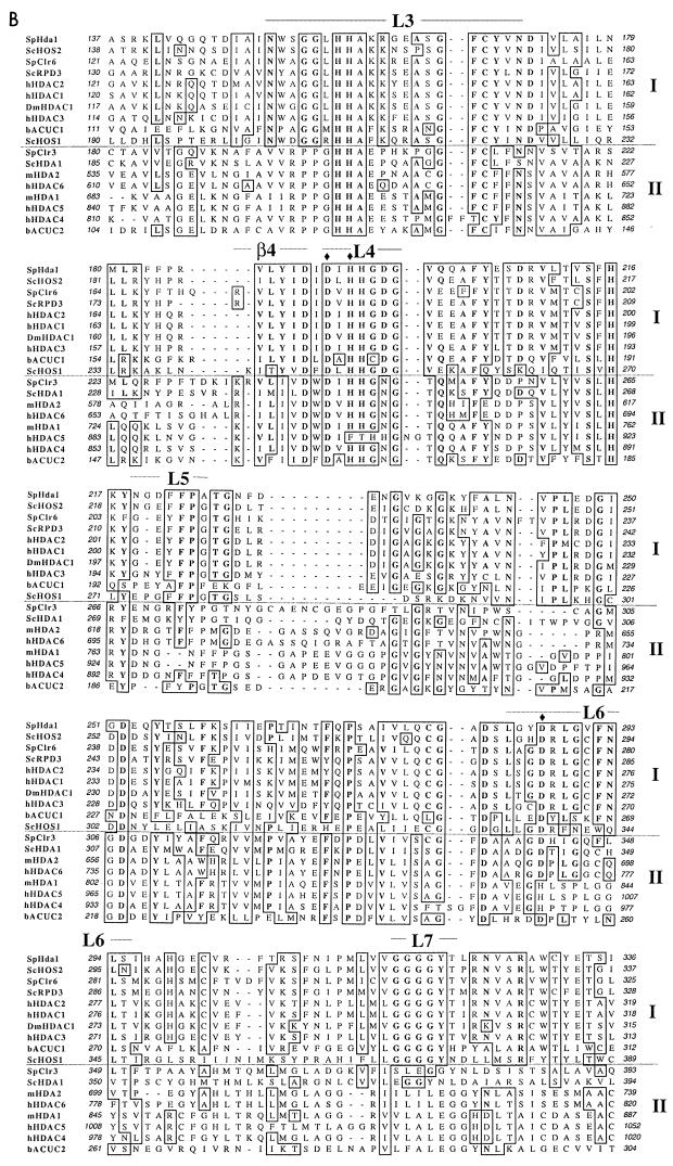
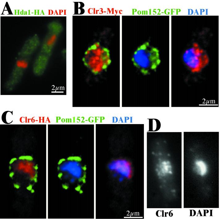
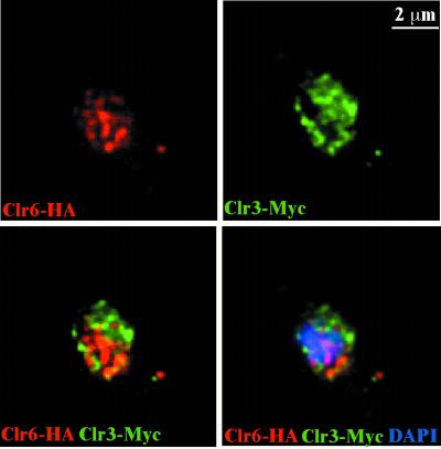
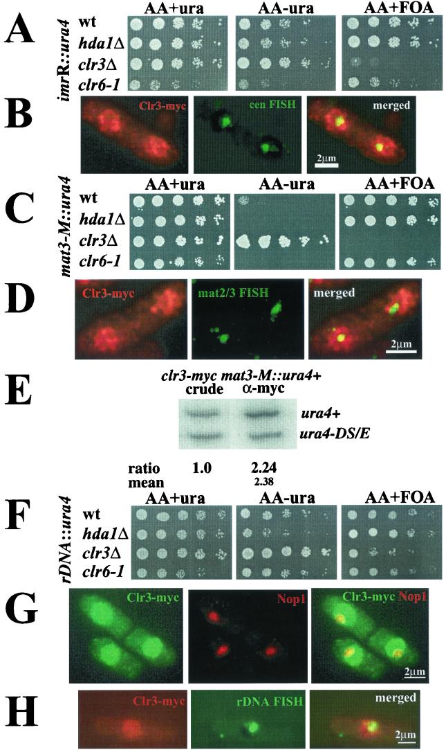
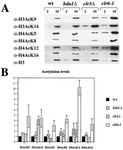
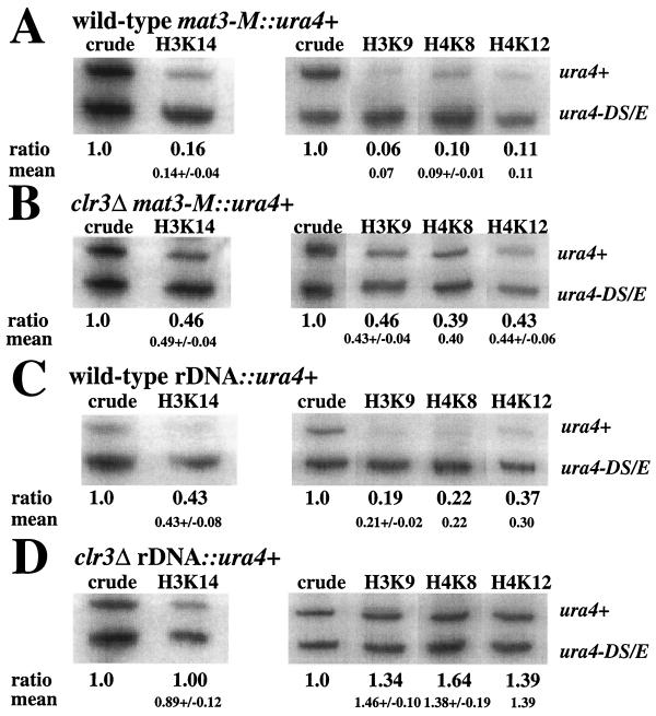
Histone deacetylases (HDACs) are important for gene regulation and the maintenance of heterochromatin in eukaryotes. Schizosaccharomyces pombe was used as a model system to investigate the functional divergence within this conserved enzyme family. S. pombe has three HDACs encoded by the hda1(+), clr3(+), and clr6(+) genes. Strains mutated in these genes have previously been shown to display strikingly different phenotypes when assayed for viability, chromosome loss, and silencing. Here, conserved differences in the substrate binding pocket identify Clr6 and Hda1 as class I HDACs, while Clr3 belongs in the class II family. Furthermore, these HDACs were shown to have strikingly different subcellular localization patterns. Hda1 was localized to the cytoplasm, while most of Clr3 resided throughout the nucleus. Finally, Clr6 was localized exclusively on the chromosomes in a spotted pattern. Interestingly, Clr3, the only HDAC present in the nucleolus, was required for ribosomal DNA (rDNA) silencing. Clr3 presumably acts directly on heterochromatin, since it colocalized with the centromere, mating-type region, and rDNA as visualized by in situ hybridization. In addition, Clr3 could be cross-linked to mat3 in chromatin immunoprecipitation experiments. Western analysis of bulk histone preparations indicated that Hda1 (class I) had a generally low level of activity in vivo and Clr6 (class I) had a high level of activity and broad in vivo substrate specificity, whereas Clr3 (class II) displayed its main activity on acetylated lysine 14 of histone H3. Thus, the distinct functions of the S. pombe HDACs are likely explained by their distinct cellular localization and their different in vivo specificities.


Figures

FIG. 1.
FIG. 1.
S. pombe HDACs are members of both phylogenetic classes. (A) A dendrogram representing the phylogenetic relationships of the HDAC family was constructed by ClustalW alignment (see Materials and Methods). The species is indicated immediately before the protein name: for example, ScHDA1 (S. cerevisiae HDA1). Abbreviations: Hs, H. sapiens; Dm, D. melanogaster; b, A. aeolicus; Sc, S. cerevisiae; Sp, S. pombe; m, Mus musculus. The two phylogenetic classes are indicated in the figure. (B) Part of the sequence alignment showing the enzymatic core region, which is the conserved part of the HDAC proteins. Class I and class II are indicated. L3 to L7 are the loops that are known to form a pocket domain around the active site. The amino acid residues coordinating the Zn2+ ion in the catalytic site are indicated (solid diamonds). β4 is a β-sheet stretch in close proximity to the active pocket.
FIG. 1.
FIG. 1.
S. pombe HDACs are members of both phylogenetic classes. (A) A dendrogram representing the phylogenetic relationships of the HDAC family was constructed by ClustalW alignment (see Materials and Methods). The species is indicated immediately before the protein name: for example, ScHDA1 (S. cerevisiae HDA1). Abbreviations: Hs, H. sapiens; Dm, D. melanogaster; b, A. aeolicus; Sc, S. cerevisiae; Sp, S. pombe; m, Mus musculus. The two phylogenetic classes are indicated in the figure. (B) Part of the sequence alignment showing the enzymatic core region, which is the conserved part of the HDAC proteins. Class I and class II are indicated. L3 to L7 are the loops that are known to form a pocket domain around the active site. The amino acid residues coordinating the Zn2+ ion in the catalytic site are indicated (solid diamonds). β4 is a β-sheet stretch in close proximity to the active pocket.
FIG. 2.
FIG. 2.
Each of the HDACs in S. pombe has a distinct characteristic cellular localization pattern. Cells of the indicated strains were processed for immunofluorescence microscopy. (A) The strain Hu268 (hda1-HA) was stained with DAPI (red) and HA antibodies (green). Hda1-HA (green) is located primarily in the cytoplasm. (B) The strain Hu397 (clr3-myc pom152-GFP) was stained with anti-myc (red) and anti-GFP (green) antibodies and DAPI (blue). The Clr3-Myc pattern colocalizes with the DNA (blue), but is not restricted to the chromatin-containing regions of the nucleus. (C) The strain Hu401 (clr6-HA pom152-GFP) was stained with anti-HA (red) and anti-GFP (green) antibodies and DAPI (blue). The Clr6-HA (red) foci were restricted to the chromatin and thus excluded from the perinuclear region, surrounded by the Pom152-GFP counter stain. (D) The strain FY648 (wild type) was stained with anti-Clr6 antibodies and DAPI. (B, C, and D) The immunofluorescence microscopy images were deconvolved (z = 0.3 μm) and merged.
FIG. 3.
FIG. 3.
Clr6 and Clr3 nuclear foci do not coincide. The strain Hu067 (clr6-HA clr3-myc) was processed for immunofluorescence microscopy and stained with anti-HA (red) and anti-myc (green) antibodies and DAPI (blue). The spotted nuclear patterns of the Clr6-HA (red) and Clr3-Myc (green) rarely colocalized completely (yellow) after deconvolution (z = 0.3 μm).
FIG.4.
FIG.4.
Clr3 is necessary for silencing of the mating-type region and the rDNA. (A, C, and F) Silencing assays. Cell suspensions were fivefold serially diluted, and each dilution was spotted onto the indicated medium and incubated for 3 days at 30°C. (A) The strains FY498 (wild type [wt]), Hu383 (hda1Δ), Hu379 (clr3Δ), and SPG141 (clr6-1) have the ura4+ gene inserted into the outer repeats of the centromere [imrR(NcoI)::ura4+]. (C) The strains FY597 (wt), FY2606 (hda1Δ), Hu427 (clr3Δ), and Hu460 (clr6-1) have the ura4+ gene inserted next to the mating-type region (mat3-M::ura4+). (F) The strains Hu393 (wt), Hu434 (hda1Δ), Hu395 (clr3Δ), and Hu451 (clr6-1) have the ura4+ gene inserted into the rDNA repeats (rDNA::ura4+). (B, D, and H) FISH experiments with Clr3-myc. Clr3-myc were processed for immunofluorescence microscopy and subjected to FISH. (B) The pRS314 cosmid was used to detect the centromere (green); Clr3-myc appears red. (D) The c1555 probe was used to detect the mat2/3 region (green); Clr3-myc appears red. (H) The rDNA probe was used for FISH (green); Clr3-myc appears red. The pictures were deconvolved (z = 0.2 μm) and merged. Yellow color indicates colocalization. (E) ChIP of Clr3-myc to the mat3-M::ura4+ marker. The strain Hu619 was subjected to ChIP. (G) Hu56 cells were stained against Clr3-myc (green) and (G) Nop1 (red). Pictures were deconvolved (z = 0.2 μm) and merged.
FIG. 5.
FIG. 5.
The acetylation pattern in bulk histone preparations of the wild type and HDAC mutants. (A) Two and 10 μg of crude histone preparations from the strains FY498 (wild type [wt]), Hu383 (hda1Δ), Hu379 (clr3Δ), and SPG141 (clr6-1) were separated by SDS-PAGE, blotted and the blots were incubated with antibodies specific for the histone variants indicated to the left of the picture. At the bottom, a Western blot with an antibody against the C terminus of histone H3 is shown as a loading control. (B) The mean and range of the intensity of the bands in two to three experiments are shown.
FIG. 6.
FIG. 6.
clr3Δ cells exhibit elevated acetylation levels at several lysines in H3 and H4 in the mating-type region and at the rDNA regions. DNA from crude chromatin preparation (crude) and immunoprecipitated (ChIP) chromatin of the indicated strains was amplified by radioactive PCR. The normalized ratios between the two variants of the ura4+ gene (ura4+ and ura4-DS/E) are shown below each lane, and the mean value of duplicate or triplicate experiments is shown underneath. The full-length ura4+ gene was either inserted (A) next to the mating-type region (mat3-M::ura4+) in strains FY597 (wild type) and (B) Hu427 (clr3Δ) or (C) in the rDNA (rDNA::ura4+) in strains Hu393 (wild type) and (D) Hu395 (clr3Δ).

References

    1. Allshire, R. C., E. R. Nimmo, K. Ekwall, J. P. Javerzat, and G. Cranston. 1995. Mutations derepressing silent centromeric domains in fission yeast disrupt chromosome segregation. Genes Dev. 9:218-233. - PubMed
    1. Bahler, J., J. Q. Wu, M. S. Longtine, N. G. Shah, A. McKenzie III, A. B. Steever, A. Wach, P. Philippsen, and J. R. Pringle. 1998. Heterologous modules for efficient and versatile PCR-based gene targeting in Schizosaccharomyces pombe. Yeast 14:943-951. - PubMed
    1. Bernstein, B. E., J. K. Tong, and S. L. Schreiber. 2000. Genomewide studies of histone deacetylase function in yeast. Proc. Natl. Acad. Sci. USA 97:13708-13713. - PMC - PubMed
    1. Bilsland, E., M. Dahlen, and P. Sunnerhagen. 1998. Genomic disruption of six budding yeast genes gives one drastic example of phenotype strain-dependence. Yeast 14:655-664. - PubMed
    1. Brownell, J. E., J. Zhou, T. Ranalli, R. Kobayashi, D. G. Edmondson, S. Y. Roth, and C. D. Allis. 1996. Tetrahymena histone acetyltransferase A: a homolog to yeast Gcn5p linking histone acetylation to gene activation. Cell 84:843-851. - PubMed

Publication types

MeSH terms

LinkOut - more resources