. 2002 Apr;22(7):2242-54.
doi: 10.1128/MCB.22.7.2242-2254.2002.

Reversal of growth suppression by p107 via direct phosphorylation by cyclin D1/cyclin-dependent kinase 4

Affiliations

Reversal of growth suppression by p107 via direct phosphorylation by cyclin D1/cyclin-dependent kinase 4

Xiaohong Leng et al. Mol Cell Biol. 2002 Apr.

Abstract

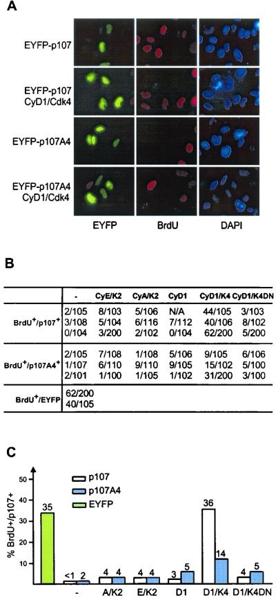
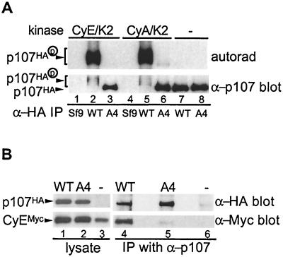
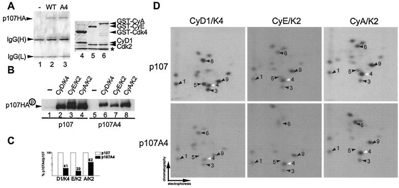
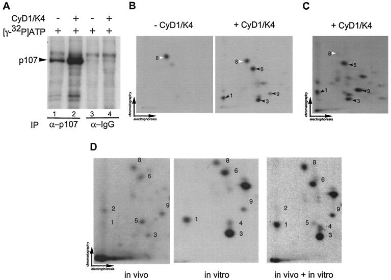
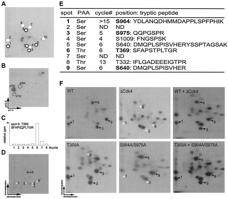
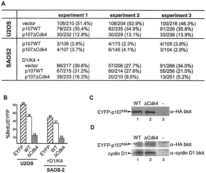
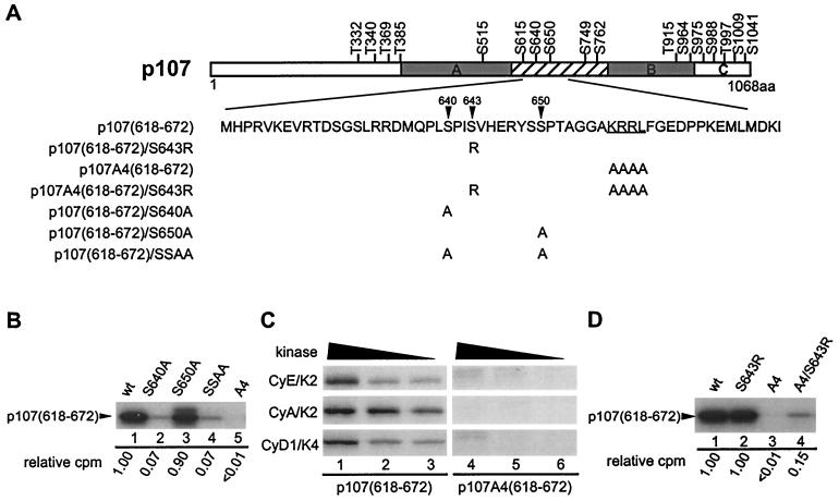
p107 functions to control cell division and development through interaction with members of the E2F family of transcription factors. p107 is phosphorylated in a cell cycle-regulated manner, and its phosphorylation leads to its release from E2F. Although it is known that p107 physically associates with E- and A-type cyclin/cyclin-dependent kinase 2 (Cdk2) complexes through a cyclin-binding RXL motif located in the spacer domain, the mechanisms underlying p107 inactivation via phosphorylation remain poorly defined. Recent genetic evidence indicates a requirement for cyclin D1/Cdk4 complexes in p107 inactivation. In this work, we provide direct biochemical evidence for the involvement of cyclin D1/Cdk4 in the inactivation of p107's growth-suppressive function. While coexpression of cyclin D1/Cdk4 can reverse the cell cycle arrest properties of p107 in Saos-2 cells, we find that p107 in which the Lys-Arg-Arg-Leu sequence of the RXL motif is replaced by four alanine residues is largely refractory to inactivation by cyclin D/Cdk4, indicating a role for this motif in p107 inactivation without a requirement for its tight interaction with cyclin D1/Cdk4. We identified four phosphorylation sites in p107 (Thr-369, Ser-640, Ser-964, and Ser-975) that are efficiently phosphorylated by Cdk4 but not by Cdk2 in vitro and are also phosphorylated in tissue culture cells. Growth suppression by p107 containing nonphosphorylatable residues in these four sites is not reversed by coexpression of cyclin D1/Cdk4. In model p107 spacer region peptides, phosphorylation of S640 by cyclin D1/Cdk4 is strictly dependent upon an intact RXL motif, but phosphorylation of this site in the absence of an RXL motif can be partially restored by replacement of S643 by arginine. This suggests that one role for the RXL motif is to facilitate phosphorylation of nonconsensus Cdk substrates. Taken together, these data indicate that p107 is inactivated by cyclin D1/Cdk4 via direct phosphorylation and that the RXL motif of p107 plays a role in its inactivation by Cdk4 in the absence of stable binding.


Figures

FIG. 1.
FIG. 1.
The Cdk interacting surface and the hydrophobic patch represent the major surfaces conserved in A-, E-, and D-type cyclins. Multisequence alignments for 17 vertebrate A-, D-, and E-type cyclins were generated and used to project on the structure of cyclin A. Residues that are conserved in character are shown in white. Residues that are not conserved among all three cyclins appear in purple. (A) Face of the projection containing the hydrophobic surface that interacts with RXL motifs. (B) Face of the projection containing the Cdk interacting surface. (C) Pairwise surface representation of the hydrophobic pocket bound to the RXL motif of p27 (stick model, backbone in yellow, side chains in gray). Residues conserved in all three proteins are shown in white. Residues conserved in cyclins A and D are shown in red, residues conserved in cyclins D and E are shown in green, and residues conserved in cyclins A and E are shown in blue. Surface residues that are not conserved are shown in gray. (D) Pairwise surface representation of the Cdk interaction surface in the composite cyclin. The PSTAIRE helix in Cdk2 is modeled onto the cyclin surface and is shown in the pink ribbon trace. Color coding is as described for panel C.
FIG. 2.
FIG. 2.
The cyclin-binding RXL motif in the spacer of p107 is dispensable for p107-induced growth arrest, but it is required for full inactivation of p107-mediated growth suppression by cyclin D1/Cdk4. (A) Saos-2 cells were transfected with vectors expressing EYFP-p107 or EYFP-p107A4 (2 μg) alone or together with the indicated cyclin/Cdk (1 μg each). At 24 h after transfection, cells were labeled with BrdU for 6 h before immunostaining. p107 expression was visualized as EYFP fluorescence. BrdU incorporation was detected by using anti-BrdU antibodies (red). Nuclei were stained with DAPI (blue). (B) Determination of the number of EYFP-p107 positive cells that are also BrdU positive from multiple experiments. (C) Summary of results in panel B depicting the proportions of p107-expressing cells that incorporated BrdU.
FIG. 3.
FIG. 3.
The RXL motif in the spacer of p107 is required for stable interaction with cyclin E and cyclin A. (A) A total of 500 ng of immunopurified p107HA and p107A4HA (in which the KRRL motif is replaced by AAAA) was incubated with purified GST-cyclin E/Cdk2 or GST-cyclin A/Cdk2 (limiting subunit, ∼500 ng) for 2 h at 4°C to allow for complex assembly. Free Cdk complexes were removed by washing complexes with binding buffer, and the remaining complexes were washed with kinase buffer. Complexes were incubated with [γ-32P]ATP (5 μCi) and ATP (50 μM) at 37°C for 30 min, followed by SDS-8% PAGE. The proteins were transferred to NC membrane, followed by autoradiography and Western blotting. (B) pcDNA3-p107HA or p107A4HA (5 μg) were cotransfected with vectors expressing Myc-tagged cyclin E (cyclin EMyc) and Cdk2 (0.5 μg each) into U2OS cells, and the cell lysates were subjected to immunoprecipitation with anti-p107 antibodies. Immune complexes and crude lysates were fractionated by SDS-8 to 16% PAGE, followed by Western blotting with anti-HA and anti-Myc antibodies.
FIG. 4.
FIG. 4.
Selective phosphorylation of recombinant p107 by cyclin D1/Cdk4 and Cdk2 kinase complexes in vitro. (A) Purified proteins used in this study. p107HA and p107A4HA were produced in insect cells and purified by immunoaffinity chromatography by using anti-HA antibodies. Proteins were separated by SDS-PAGE and stained with Coomassie blue (lanes 2 and 3). GST-cyclin A/Cdk2 (lane 6), GST-cyclin E/Cdk2 (lane 5), and GST-cyclin D1/Cdk4 (lane 4) were purified as described previously (15) and subjected to SDS-PAGE prior to staining with Coomassie blue. The asterisk indicates the position of insect cell GST which copurifies with the recombinant GST proteins. (B) Immunopurified p107HA and p107A4HA (500 nM) were subjected to in vitro kinase assays with purified cyclin D1/GST-Cdk4, GST-cyclin E/Cdk2, and GST-cyclin A/Cdk2 (20 nM) without a prebinding step. Reaction products were fractionated by SDS-8% PAGE and transferred to nitrocellulose; 32P-incorporation was then visualized by autoradiography. (C) Normalization of 32P incorporation from panel B. (D) Two-dimensional tryptic phosphopeptide mapping of phosphorylated p107HA or p107A4HA derived from panel B. Spot 4 (white arrow) is preferentially phosphorylated by Cdk2 complexes relative to Cdk4 complexes. Phosphopeptides 1, 3, 6, and 9 are preferentially phosphorylated by Cdk4. Among these phosphopeptides, spot 9 is also prominent in Cdk2 peptide maps.
FIG. 5.
FIG. 5.
Visualization of cyclin D1/Cdk4-specific phosphorylation events in p107. (A) Anti-p107 or control immune complexes from asynchronous C33A cells were incubated with [γ-32P]ATP in the presence or absence of purified cyclin D1/Cdk4 (40 nM). Reaction mixtures were separated by SDS-8% PAGE and transferred to nitrocellulose prior to autoradiography. (B) Two-dimensional tryptic phosphopeptide mapping of samples derived from panel A. In the absence of Cdk4, the single major phosphopeptide 8 was found (white arrow). In the presence of Cdk4, four additional phosphopeptides (peptides 1, 3, 6, and 9) were observed (black arrow). (C) Phosphorylation of p107 immune complexes from Saos-2 cells by cyclin D1/Cdk4 in vitro. pcDNA-p107HA was transiently expressed in Saos-2 cells, and anti-HA immune complexes were then used in kinase assays with cyclin D1/Cdk4 as described in panel B. Phosphorylated p107HA was subjected to tryptic peptide mapping. The major phosphopeptides seen with Cdk4 in panel B were found in panel C (peptides 1, 3, 6, and 9) as indicated by the black arrows. Peptide 8 (white arrow) was weakly phosphorylated in this setting. (D) In vivo phosphorylation of p107. p107HA was expressed in U2OS cells by transient transfection prior to metabolic labeling and analysis as described in Materials and Methods. Labeled p107 (600 cpm) was subjected to peptide mapping in the presence or absence of in vitro-phosphorylated p107HA obtained by immunoprecipitation from U2OS cells.
FIG. 6.
FIG. 6.
Determination of in vitro Cdk phosphorylation sites in p107. (A) Designation of p107 phosphopeptides. Major phosphopeptides (designated 1 to 10) are indicated schematically with open circles for Cdk2 preferred sites and with closed circles for Cdk4 preferred sites. (B) Phosphoamino acid analysis of phosphopeptide 6. Tryptic phosphopeptide 6 from Cdk4 phosphorylated p107 (Fig. 4D) was eluted from a two-dimensional electrophoresis plate and subjected to phosphoamino acid analysis. The positions of unlabeled phosphoamino acids used as standards are circled. The phosphorylated amino acid was visualized by autoradiography. (C) Edman degradation of phosphopeptide 6. Residues released at each Edman cycle were subjected to liquid scintillation counting, and the radioactivity at each cycle is presented as a histogram. Based on p107 tryptic peptides, phosphopeptide 6 was predicted to correspond to SFAPS TPLTGR, where the underlined residue is phosphorylated. (D) A synthetic tryptic peptide corresponding to SFAPS TPLTGR was phosphorylated in vitro with cyclin D1/Cdk4, purified, and then subjected to peptide mapping with a limiting amount of p107HA previously phosphorylated by cyclin D1/Cdk4. The phosphorylated synthetic peptide comigrated with phosphopeptide 6 from p107, as indicated by the solid arrow. (E) Summary of Cdk phosphorylation sites in p107 identified in this study. Cyclin D1/Cdk4-specific sites are indicated in boldface type. (F) Major cyclin D1/Cdk4 phosphorylation sites are absent in the p107ΔCdk4 mutant in which T369, S640, S964, and S975 are replaced by alanine. HA-tagged p107, p107(T369A), p107(S964A/S975A), and p107ΔCdk4 were expressed in U2OS cells, purified by using anti-HA antibodies, and phosphorylated by cyclin D1/Cdk4 (40 nM) in vitro. Proteins were subjected to two-dimensional peptide mapping either alone or in various combinations. The positions of Cdk4 phosphorylation sites are shown by black arrows when present, and their expected positions are shown by white arrows in maps derived from mutants containing nonphosphorylatable residues.
FIG. 7.
FIG. 7.
Mutation of Cdk4 phosphorylation sites in p107 abolishes the ability of cyclin D1/Cdk4 to reverse growth suppression. (A) p107 insensitive U2OS cells or p107-sensitive Saos-2 cells were transfected with vectors expressing EYFP-p107 or EYFP-p107ΔCdk4 (3 μg) in the presence or absence of vectors expressing cyclin D1 and Cdk4 (1 μg each). After 24 h, cells were labeled with BrdU. EYFP-p107-positive cells incorporating BrdU were determined by immunofluorescence. The numbers of BrdU-positive and EYFP-p107-positive cells are shown for multiple independent experiments. (B) Histogram of data presented in panel A. (C) The levels of p107 and p107ΔCdk4 expressed in U2OS cells were similar, as determined by immunoblotting with anti-p107 antibodies. (D) Immunoblots of cyclin D1 and EYFP-p107 proteins revealed similar expression levels in Saos-2 cells.
FIG. 8.
FIG. 8.
Phosphorylation of S640 is RXL dependent in p107(618-672). (A) Schematic representation of spacer mutants used in this study. (B) S640 is the Cdk target site in p107(618-672). The indicated proteins were phosphorylated by cyclin A/Cdk2 (20 nM) in vitro. Reaction products were separated on 8 to 20% Tricine gels, followed by autoradiography and quantitation with a PhosphorImager. (C) Phosphorylation of S640 is RXL dependent. GST-p107(618-672) and GST-p107A4(618-672) were incubated with the indicated kinases and [γ-32P]ATP, and the reaction products were separated by SDS-PAGE prior to autoradiography. (D) Cdk phosphorylation on S640 is abolished in the absence of RXL motif, but it is partially restored by replacing the +3 residue with arginine. Reactions were performed as in panel B. Quantitation by PhosphorImager analysis is indicated below each lane.
FIG. 9.
FIG. 9.
(A) Conserved organization of Cdk4 phosphorylation sites in pocket proteins. Major Cdk4 phosphorylation sites are indicated by black arrows. The position of the RXL motif in p107 and p130 is indicated by an asterisk. (B) Alignment of Cdk4 phosphorylation sites in p107 (the present study), p130 (21), and Rb (15, 30, 64).

Similar articles

Cited by

References

    1. Adams, P. D., and J. W. Harper. 2001. Cyclin-dependent kinases. Chem. Rev. 101:2511-2526. - PubMed
    1. Adams, P. D., W. R. Sellers, S. K. Sharma, A. D. Wu, C. M. Nalin, and W. G. Kaelin, Jr. 1996. Identification of a cyclin-Cdk2 recognition motif present in substrates and p21-like cyclin-dependent kinase inhibitors. Mol. Cell. Biol. 16:6623-6633. - PMC - PubMed
    1. Adams, P. D., X. Li, W. R. Sellers, K. B. Baker, X. Leng, J. W. Harper, Y. Taya, and W. G. Kaelin, Jr. 1999. Retinoblastoma protein contains a C-terminal motif that targets it for phosphorylation by cyclin-Cdk complexes. Mol. Cell. Biol. 19:1068-1080. - PMC - PubMed
    1. Aktas, H., H. Cai, and G. M. Cooper. 1997. Ras links growth factor signaling to the cell cycle machinery via regulation of cyclin D1 and the Cdk inhibitor p27KIP1. Mol. Cell. Biol. 17:3850-3857. - PMC - PubMed
    1. Alevizopoulos, K., J. Vlach, S. Hennecke, and B. Amati. 1997. Cyclin E and c-Myc promote cell proliferation in the presence of p16INK4a and of hypophosphorylated retinoblastoma family proteins. EMBO J. 16:5322-5333. - PMC - PubMed

Publication types

MeSH terms