. 2002 Apr;22(7):2294-303.
doi: 10.1128/MCB.22.7.2294-2303.2002.

The ubiquitin ligase component Siah1a is required for completion of meiosis I in male mice

Affiliations

The ubiquitin ligase component Siah1a is required for completion of meiosis I in male mice

Ross A Dickins et al. Mol Cell Biol. 2002 Apr.

Abstract

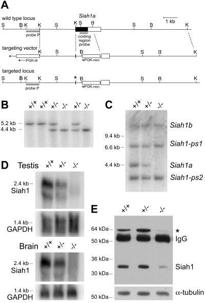
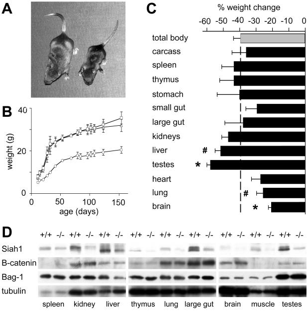
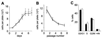
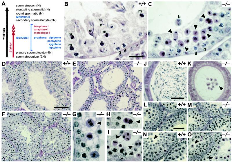
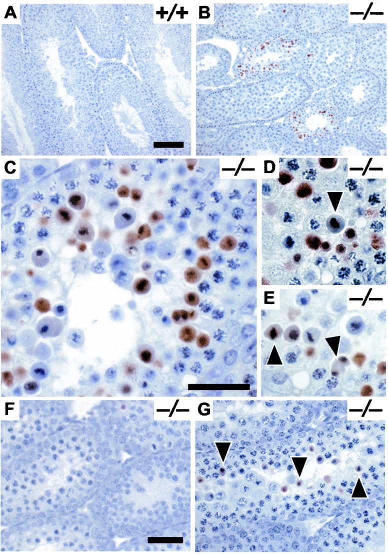
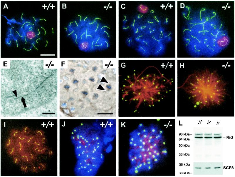
The mammalian Siah genes encode highly conserved proteins containing a RING domain. As components of E3 ubiquitin ligase complexes, Siah proteins facilitate the ubiquitination and degradation of diverse protein partners including beta-catenin, N-CoR, and DCC. We used gene targeting in mice to analyze the function of Siah1a during mammalian development and reveal novel roles in growth, viability, and fertility. Mutant animals have normal weights at term but are postnatally growth retarded, despite normal levels of pituitary growth hormone. Embryonic fibroblasts isolated from mutant animals grow normally. Most animals die before weaning, and few survive beyond 3 months. Serum gonadotropin levels are normal in Siah1a mutant mice; however, females are subfertile and males are sterile due to a block in spermatogenesis. Although spermatocytes in mutant mice display normal meiotic prophase and meiosis I spindle formation, they accumulate at metaphase to telophase of meiosis I and subsequently undergo apoptosis. The requirement of Siah1a for normal progression beyond metaphase I suggests that Siah1a may be part of a novel E3 complex acting late in the first meiotic division.


Figures

FIG. 1.
FIG. 1.
Disruption of Siah1a by gene targeting. (A) Targeting strategy Box, single coding exon of the wild-type Siah1a locus (coding region is black). The targeting vector is shown below the wild-type locus, with parallel dashed lines bordering the targeting sequences. Homologous recombination yields the targeted locus, containing only the first 22 codons of the Siah1a coding region (∗). B, BstXI; K, KpnI; S, SacI. (B) Southern analysis of BstXI-digested genomic DNA from progeny of a Siah1a+/− intercross. Probe P hybridizes to a region 5′ of the targeting sequence and was used to distinguish between the wild-type (5.2-kb) and targeted (4.4-kb) alleles. (C) Southern analysis of SacI-digested genomic DNA from progeny of a Siah1a+/− intercross, probed with a coding region fragment that hybridizes to the four murine Siah1 genes including pseudogenes Siah1-ps1 and Siah1-ps2. (D) Northern blot of mRNA isolated from testes and brains of mutant and control mice and hybridized with the Siah1 coding region fragment used for Southern analysis. Blots were stripped and reprobed with a GAPDH (glyceraldehyde-3-phosphate dehydrogenase) loading control. (E) Western blot of whole-testis lysates from mutant and control mice, blotted with anti-Siah1 monoclonal antibody 3A9. ∗, 64-kDa species.
FIG. 2.
FIG. 2.
Siah1a−/− mice are growth retarded. (A) Small size of a 2-week-old Siah1a mutant mouse (right) compared to that of a sex-matched wild-type littermate (left). (B) Representative growth curve (means and ranges) of male Siah1a+/+ (squares; n = 2), Siah1a+/− (triangles; n = 4), and Siah1a−/− (circles; n = 2) mice from two age-matched litters. Data for females showed a similar trend. (C) Wet weights (means ± standard errors of the means) of tissues from Siah1a−/− animals expressed as percent changes compared to those of wild-type littermates. Data were obtained from mice between 3 and 10 weeks of age (n = 7 to 10; n = 16 for testes). ∗, P < 0.001; #, P < 0.05 (both compared to change in total body weight (grey). (D) Western blot of organ lysates from mutant and control mice, blotted with antibodies recognizing Siah1, β-catenin, and Bag-1.
FIG. 3.
FIG. 3.
Growth properties of Siah1a−/− MEFs. Means ± standard deviations (Siah1a+/+embryos, n = 2; Siah1a−/− embryos, n = 3) from duplicate assays of each of n independent MEF preparations derived from littermate e14 embryos are shown. Similar results were obtained with MEFs derived from separate litters. Black, wild type; grey (A and B) or white (C), Siah1a−/−. (A) MEF growth. Passage 4 MEFs were plated at 2 × 10⁵ cells/60-mm-diameter culture dish, and cell numbers were determined daily for 7 days. (B) 3T3 analysis of MEF proliferation and senescence. Passage 4 MEFs were plated at 3 × 10⁵ cells/60-mm-diameter culture dish. Cell numbers were determined after 3 days, before replating at the starting density. MEFs of both genotypes underwent senescence by passage 7. (C) Cell cycle distribution of asynchronously growing passage 4 MEFs. Flow cytometry using 5-bromo-2′-deoxyuridine labeling and propidium iodide staining was used to assess the percentage of cells in each phase of the cell cycle.
FIG. 4.
FIG. 4.
Defective spermatogenesis in Siah1a−/− mice. (A) Schematic of spermatogenesis, including the stages of meiosis I where Siah1a−/− spermatogenesis is clearly aberrant (red). Not all cell types listed are present in a single seminiferous tubule section. (B to O) Hematoxylin- and eosin-stained testis and epididymis sections from representative Siah1a−/− and control mice. (B to I) Testis sections from adult mice. (B) Wild type at stage XII (40), showing spermatogonia (g), metaphase I spermatocytes (m), and elongating spermatids (e). (C) Severely affected Siah1a−/− testis at a stage similar to that shown in panel B but with clearly abnormal dividing spermatocytes at metaphase through telophase I (arrowheads). Note the abnormally eosinophilic cytoplasm of degenerating dividing spermatocytes, a binucleated spermatid (b), and the absence of elongating spermatids. (D to F) Normal testis morphology of wild-type mice (D) compared to the complete or partial absence of postmeiotic cells in mutants with severe (E) or mild (F) phenotypes. (G to I) Abnormal spermatocytes in mutant animals. Defects observed include apparently degenerating metaphase and anaphase I spermatocytes (G and H; arrowheads) and unusual chromatin figures and binucleated cells (I, arrowheads). (J and K) Epididymis sections showing abundant spermatozoa in the wild type (J) compared to mutant (K), which contains only immature spermatocytes. Many of these have chromatin figures reminiscent of meiotically dividing cells (arrowhead). (L to O) Testis sections from wild-type (L and N) and mutant (M and O) juvenile mice. At day 18 (L and M) the wild type and mutant are comparable. By day 22 (N and O) round spermatids are present in the wild type (arrowhead); however Siah1a−/− tubules harbor abnormal spermatocytes undergoing meiotic division (arrowhead). Bars: 10 (B, C, and G to I), 50 (D to F), and 20 μm (J to O).
FIG. 5.
FIG. 5.
In situ staining of apoptotic spermatocytes in mutant testes. DNA fragmentation was visualized by TUNEL staining (hematoxylin counterstained). (A to E) Adult testes from wild-type (A) and mutant (B to E) mice. Apoptosis in mutants is confined to particular seminiferous tubules (B), all containing meiotically dividing spermatocytes (C). Apoptotic DNA fragmentation (D and E) is clearly visible in spermatocytes at metaphase I to telophase I (arrowheads). (F and G) Developmental analysis of apoptosis in testis from Siah1a−/− mice at days 18 (F) and 22 (G). Note the appearance of numerous apoptotic spermatocytes (arrowheads) at day 22. Bars: 50 (A and B), 10 (C to E), and 20 μm (F and G).
FIG. 6.
FIG. 6.
Meiotic prophase analysis and Kid expression in mutant spermatocytes. Thousands of spermatocytes from several wild-type and Siah1a−/− mice exhibiting various degrees of phenotypic severity were examined. Typical cells are shown. (A to D) Spermatocytes from wild type (A and C) and Siah1a−/− (B and D) germ cell spreads stained with DAPI to visualize DNA (blue) and antibodies to SCP3 (green) and SUMO-1 (red). SUMO-1 localizes to the sex body, allowing identification of the paired X and Y chromosomes. (A and B) Pachytene spermatocytes showing 19 fully synapsed autosomal bivalents and an X-Y chromosome pair. (C and D) Diplotene spermatocytes showing normal chiasma formation and end-to-end X-Y chromosome attachment. (E) Transmission electron micrograph of a Siah1a−/− pachytene spermatocyte. The synaptonemal complex is morphologically normal; lateral (arrow) and central (arrowhead) elements are indicated. (F) Anti-α-tubulin immunohistochemistry showing normal spindle morphology (arrowheads) in Siah1a−/− metaphase I spermatocytes. (G and H) Metaphase I spermatocytes from wild-type (G) and Siah1a mutant (H) mice stained with DAPI and antibodies to SCP3 (green) and α-tubulin (red). Each spermatocyte has settled down flat, collapsing on its spindle pole axis. Thus bipolar spindle morphology is not seen but kinetochores are easily distinguishable. Each of the 40 SCP3 foci represents a superimposed pair of sister kinetochores (6), corresponding to the diploid chromosome number. Kinetochore attachment and spindle morphology in wild-type spermatocytes are comparable to those in Siah1a−/− spermatocytes. (I to L) Kid expression in mammalian spermatocytes. (I to K) Spermatocytes from wild-type (I and J) and Siah1a−/− (K) germ cell spreads stained with antibodies to SCP3 (green) and Kid (red). Metaphase I spermatocytes (J and K) are also stained with DAPI (blue). Kid localizes to chromatin in prophase spermatocytes (I) and to the metaphase I spindle in spermatocytes of both genotypes (J and K). (L) Western blot of whole-testis lysates from mutant and control mice, blotted with the anti-Kid antibody. Kid migrates at approximately 70 kDa. Bars: 5 (A to D, G, H, and I to K), 1 (E), and 10 μm (F).

References

    1. Amson, R. B., M. Nemani, J. P. Roperch, D. Israeli, L. Bougueleret, I. Le Gall, M. Medhioub, G. Linares-Cruz, F. Lethrosne, P. Pasturaud, L. Piouffre, S. Prieur, et al. 1996. Isolation of 10 differentially expressed cDNAs in p53-induced apoptosis: activation of the vertebrate homologue of the Drosophila seven in absentia gene. Proc. Natl. Acad. Sci. USA 93:3953-3957. - PMC - PubMed
    1. Antonio, C., I. Ferby, H. Wilhelm, M. Jones, E. Karsenti, A. R. Nebreda, and I. Vernos. 2000. Xkid, a chromokinesin required for chromosome alignment on the metaphase plate. Cell 102:425-435. - PubMed
    1. Baker, S. M., A. W. Plug, T. A. Prolla, C. E. Bronner, A. C. Harris, X. Yao, D. M. Christie, C. Monell, N. Arnheim, A. Bradley, T. Ashley, and R. M. Liskay. 1996. Involvement of mouse Mlh1 in DNA mismatch repair and meiotic crossing over. Nat. Genet. 13:336-342. - PubMed
    1. Boehm, J., Y. He, A. Greiner, L. Staudt, and T. Wirth. 2001. Regulation of BOB.1/OBF.1 stability by SIAH. EMBO J. 20:4153-4162. - PMC - PubMed
    1. Boulton, S. J., A. Brook, K. Staehling-Hampton, P. Heitzler, and N. Dyson. 2000. A role for Ebi in neuronal cell cycle control. EMBO J. 19:5376-5386. - PMC - PubMed

Publication types

MeSH terms