. 2002 Apr;22(7):2375-87.
doi: 10.1128/MCB.22.7.2375-2387.2002.

Protein phosphatase 2A forms a molecular complex with Shc and regulates Shc tyrosine phosphorylation and downstream mitogenic signaling

Affiliations

Protein phosphatase 2A forms a molecular complex with Shc and regulates Shc tyrosine phosphorylation and downstream mitogenic signaling

Satoshi Ugi et al. Mol Cell Biol. 2002 Apr.

Abstract

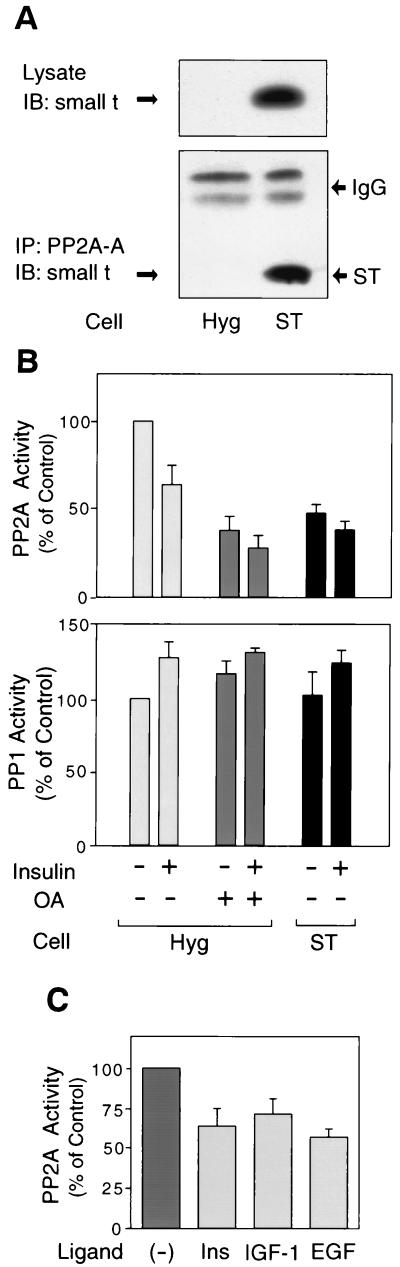
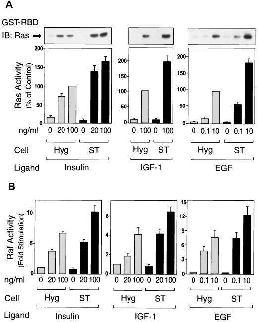
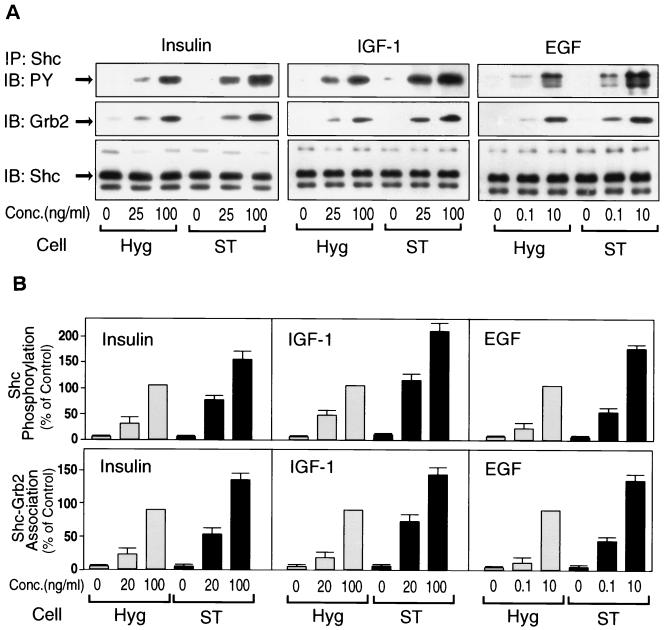
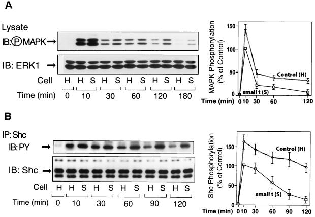
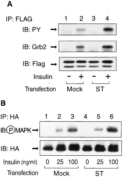
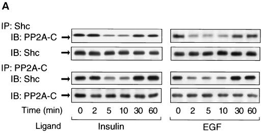
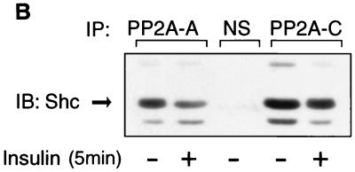
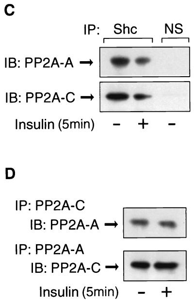
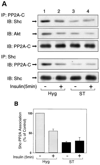
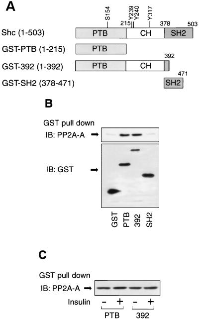
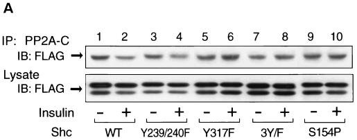
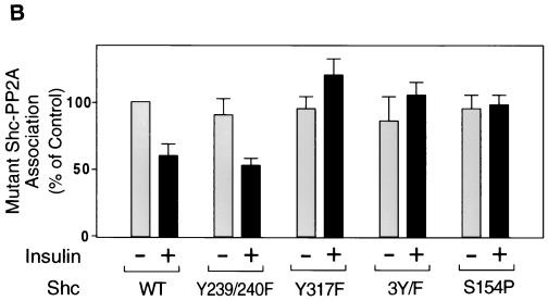
Protein phosphatase 2A (PP2A) is a multimeric serine/threonine phosphatase that carries out multiple functions. Although numerous observations suggest that PP2A plays a major role in downregulation of the mitogen-activated protein (MAP) kinase pathway, the precise mechanisms are unknown. To clarify the role of PP2A in growth factor (insulin, epidermal growth factor [EGF], and insulin-like growth factor 1 [IGF-1]) stimulation of the Ras/MAP kinase pathway, simian virus 40 small t antigen was expressed in Rat-1 fibroblasts which overexpress insulin receptors. Small t antigen is known to specifically inhibit PP2A by binding to the A PP2A regulatory subunit, interfering with the ability of PP2A to bind to its cellular substrates. Overexpressed small t protein was coimmunoprecipitated with PP2A and inhibited cellular PP2A activity but did not inhibit protein phosphatase 1 (PP1) activity. Insulin, IGF-1, and EGF stimulation also inhibited PP2A activity. Growth factor-stimulated Ras, Raf-1, MAP kinase, and mitogen-activated extracellular-signal-regulated kinase kinase (MEK) activities were elevated in small-t-antigen-expressing cells. Furthermore, Shc tyrosine phosphorylation and its association with Grb2 were also elevated in small-t-antigen-expressing cells. Expression levels of Shc, Ras, MEK, or MAP kinase and phosphorylation of insulin, EGF, and IGF-1 receptors were not altered. Interestingly, we found that PP2A associated with Shc in the basal state and dissociated in response to insulin and EGF and that this dissociation was inhibited by 65% in small-t-antigen-expressing cells. In addition, we found that PP2A associates with the phosphotyrosine-binding domain (PTB domain) of Shc and that phosphorylation of tyrosine 317 of Shc was required for PP2A-Shc dissociation. We conclude (i) that PP2A negatively regulates the Ras/MAP kinase pathway by binding to Shc, inhibiting tyrosine phosphorylation; (ii) that the Shc-PP2A association is mediated by the Shc PTB domain but the interaction is independent of phosphotyrosine binding, indicating a new molecular function for the PTB domain; (iii) that growth factor stimulation, or small-t-antigen expression, causes dissociation of the PP2A-Shc complex, facilitating Shc phosphorylation and downstream activations of the Ras/MAP kinase pathway; and (iv) that this defines a new mechanism of small-t-antigen action to promote mitogenesis.


Figures

FIG. 1.
FIG. 1.
Expression of small t inhibits PP2A activity. (A) Control cells (Hyg) and cell lines overexpressing small t (ST) were lysed, and whole-cell lysates were analyzed by Western blotting with anti-small t antibody (upper panel). The same cell lysates were immunoprecipitated with antibody against the A subunit of PP2A, followed by immunoblotting with anti-small t antibody (lower panel). (B) Starved cells were pretreated with or without 1 μM okadaic acid (OA) for 40 min and then stimulated with 100 ng of insulin/ml for 20 min. Whole-cell lysates were prepared and assayed for PP2A activity (upper panel) and PP1 activity (lower panel) as described in Materials and Methods. (C) PP2A activity was assayed in cells stimulated with 100 ng of insulin/ml for 20 min, 100 ng of IGF-1/ml for 20 min or 10 ng of EGF/ml for 15 min. Data are presented as the percentage of phosphatase activity compared to unstimulated control cells and represent the mean ± the standard error (SE) of four independent experiments.
FIG. 2.
FIG. 2.
Expression of small t enhances insulin-, IGF-1-, and EGF-stimulated Ras and Raf-1 kinase activity. Starved cells were stimulated with insulin (left panel) for 3 min, with IGF-1 (middle panel) for 3 min, or with EGF (right panel) for 1.5 min. (A) Whole-cell lysates were prepared and precipitated with the GST-RBD fusion protein. The precipitates were then analyzed by Western blotting with anti-Ras antibody as described in Materials and Methods (upper panel). Data are presented as the percentage of Ras activity when compared with maximally stimulated Hyg cells and represent the mean ± SE of three independent experiments. (B) Whole-cell lysates were prepared and assayed for Raf-1 protein kinase activity as described in Materials and Methods. Data are presented as the fold increase in Raf-1 kinase activity compared to unstimulated Hyg cells and represent the mean ± SE of four independent experiments.
FIG. 3.
FIG. 3.
Expression of small t enhances insulin-, IGF-1-, and EGF-stimulated tyrosine phosphorylation of Shc and its association with Grb2. (A) Starved cells were stimulated with insulin (left panel), IGF-1 (middle panel), or EGF (right panel) for 5 min. Whole-cell lysates were prepared and immunoprecipitated with anti-Shc antibody, followed by immunoblotting with anti-phosphotyrosine antibody (upper panel), anti-Grb2 antibody (middle panel), or anti-Shc antibody (lower panel). (B) Data are expressed as the percentage of Shc phosphorylation levels (upper panel), and its association with Grb2 (lower panel) compared with maximally stimulated Hyg cells; the results represent the mean ± SE of four independent experiments.
FIG. 4.
FIG. 4.
Expression of small t prolongs insulin-induced phosphorylation of MAP kinase and Shc. (A) Control cells (H) and cell lines overexpressing small t (S) were stimulated with insulin (100 ng/ml) for the indicated periods of time. Whole-cell lysates were analyzed by Western blotting with phospho-MAP kinase antibody (left upper panel) or anti-ERK1 antibody (left lower panel). (B) The same cell lysates were immunoprecipitated with anti-Shc antibody, followed by immunoblotting with anti-phosphotyrosine (PY) antibody (left upper panel) or anti-Shc antibody (left lower panel). Data are presented as the percentage of MAPK phosphorylation (⋄) (A, right panel) or of Shc phosphorylation (⋄) (B, right panel) compared to maximally stimulated control (□) cells and represent the mean ± SE of three independent experiments.
FIG. 5.
FIG. 5.
Transient expression of small t enhances insulin-stimulated tyrosine phosphorylation of Shc, its association with Grb2 and MAP kinase phosphorylation. (A) pRK5 vector encoding FLAG-tagged Shc was transiently cotransfected with either pCMV5 vector encoding small t (ST, lanes 3 and 4) or control vector (mock, lanes 1 and 2) in HIRc B cells as described in Materials and Methods. Cells were starved and stimulated with 100 ng of insulin/ml (lanes 2 and 4) for 5 min. Whole-cell lysates were prepared and immunoprecipitated with anti-FLAG antibody, followed by immunoblotting with anti-phosphotyrosine antibody (upper panel), anti-Grb2 antibody (middle panel), or anti-FLAG antibody (lower panel). (B) pcDNA3 vector encoding HA-tagged ERK2 was transiently cotransfected with either pCMV5 vector encoding small t (ST, lanes 4 to 6) or control vector (mock, lanes 1 to 3) in HIRc B cells. Cells were starved and stimulated with insulin for 5 min. Whole-cell lysates were prepared and immunoprecipitated with anti-HA antibody, followed by immunoblotting with anti-phospho-MAP kinase antibody (upper panel) or anti-HA antibody (lower panel).
FIG. 6.
FIG. 6.
PP2A associates with Shc and dissociates upon insulin or EGF stimulation. (A) HIRc B cells were starved and stimulated with 100 ng of insulin/ml (left panel) or 10 ng of EGF/ml (right panel) for the indicated times. Whole-cell lysates were prepared and immunoprecipitated with anti-Shc antibody, followed by immunoblotting with anti-PP2A-C antibody (top panel) or anti-Shc antibody (second panel). The same lysates were immunoprecipitated with anti-PP2A-C antibody, followed by immunoblotting with anti-Shc antibody (third panel) or anti-PP2A-C antibody (bottom panel). (B to D) Cells were stimulated with 100 ng of insulin/ml for 5 min and immunoprecipitated with anti-Shc antibody, anti-PP2A-A antibody, anti-PP2A-C antibody, or nonimmune sera (NS). The immnoprecipitates were analyzed by Western blotting with anti-Shc antibody (B), anti-PP2A-A antibody (C and D, upper panel), or anti-PP2A-C antibody (C and D, lower panel).
FIG. 6.
FIG. 6.
PP2A associates with Shc and dissociates upon insulin or EGF stimulation. (A) HIRc B cells were starved and stimulated with 100 ng of insulin/ml (left panel) or 10 ng of EGF/ml (right panel) for the indicated times. Whole-cell lysates were prepared and immunoprecipitated with anti-Shc antibody, followed by immunoblotting with anti-PP2A-C antibody (top panel) or anti-Shc antibody (second panel). The same lysates were immunoprecipitated with anti-PP2A-C antibody, followed by immunoblotting with anti-Shc antibody (third panel) or anti-PP2A-C antibody (bottom panel). (B to D) Cells were stimulated with 100 ng of insulin/ml for 5 min and immunoprecipitated with anti-Shc antibody, anti-PP2A-A antibody, anti-PP2A-C antibody, or nonimmune sera (NS). The immnoprecipitates were analyzed by Western blotting with anti-Shc antibody (B), anti-PP2A-A antibody (C and D, upper panel), or anti-PP2A-C antibody (C and D, lower panel).
FIG. 7.
FIG. 7.
Expression of small t decreases the association of PP2A with Shc. (A) Hyg cells (lanes 1 and 2) and ST cells (lanes 3 and 4) were starved and stimulated with 100 ng of insulin/ml for 5 min. Whole-cell lysates were prepared and immunoprecipitated with anti-PP2A-C antibody, followed by immunoblotting with anti-Shc antibody (top panel), anti-Akt antibody (second panel), and PP2A-C antibody (third panel). The same cell lysates were immunoprecipitated with anti-Shc antibody, followed by immunoblotting with anti-PP2A-C antibody (fourth panel) or anti-Shc antibody (bottom panel). (B) Data are presented as the percentage of the association of Shc with PP2A compared to unstimulated Hyg cells and represent the mean ± SE of four independent experiments.
FIG. 8.
FIG. 8.
Association of PP2A with GST fusion proteins containing Shc domains. (A) Schematic of Shc and constructs of GST fusion proteins. (B) PP2A associates with the PTB domain of Shc. Cell lysates from HIRc B cells were incubated with the indicated GST fusion proteins, and the precipitates were analyzed by Western blotting with anti-PP2A-A antibody as described in Materials and Methods (upper panel). The expression of GST fusion proteins were determined by Western blotting with anti-GST antibody (lower panel). (C) Cells were stimulated with or without 100 ng of insulin/ml for 5 min, and cell lysates were incubated with GST-PTB or GST-392; the precipitates were analyzed by Western blotting with anti-PP2A-A antibody.
FIG. 9.
FIG. 9.
Phosphorylation of tyrosine 317 is required for the dissociation of PP2A from Shc. (A) FLAG-tagged wild-type Shc (lanes 1 and 2), Y239/240F Shc (lanes 3 and 4), Y317F Shc (lanes 5 and 6), 3Y/F Shc (lanes 7 and 8), or S154P Shc (lanes 9 and 10) were transiently expressed in HIRc B cells as described in Materials and Methods. Cells were starved and stimulated with 100 ng of insulin/ml (lanes 2, 4, 6, 8, and 10) for 5 min. Whole-cell lysates were prepared and immunoprecipitated with anti-PP2A-C antibody, followed by immunoblotting with anti-FLAG antibody (upper panel). The same lysates were analyzed by Western blotting with anti-FLAG antibody (lower panel). (B) Data are expressed as the percentage of the Shc-PP2A association levels compared to starved cells transfected with wild-type Shc and represent the mean ± SE of three independent experiments. (C) Phosphorylation of Shc mutants. The indicated Shc mutants were transiently expressed in HIRc B cells. Cells were starved and stimulated with 100 ng of insulin/ml (lanes 2 to 6) for 5 min. Whole-cell lysates were prepared and immunoprecipitated with anti-FLAG antibody, followed by immunoblotting with antiphosphotyrosine antibody (upper panel) or anti-FLAG antibody (lower panel).
FIG. 9.
FIG. 9.
Phosphorylation of tyrosine 317 is required for the dissociation of PP2A from Shc. (A) FLAG-tagged wild-type Shc (lanes 1 and 2), Y239/240F Shc (lanes 3 and 4), Y317F Shc (lanes 5 and 6), 3Y/F Shc (lanes 7 and 8), or S154P Shc (lanes 9 and 10) were transiently expressed in HIRc B cells as described in Materials and Methods. Cells were starved and stimulated with 100 ng of insulin/ml (lanes 2, 4, 6, 8, and 10) for 5 min. Whole-cell lysates were prepared and immunoprecipitated with anti-PP2A-C antibody, followed by immunoblotting with anti-FLAG antibody (upper panel). The same lysates were analyzed by Western blotting with anti-FLAG antibody (lower panel). (B) Data are expressed as the percentage of the Shc-PP2A association levels compared to starved cells transfected with wild-type Shc and represent the mean ± SE of three independent experiments. (C) Phosphorylation of Shc mutants. The indicated Shc mutants were transiently expressed in HIRc B cells. Cells were starved and stimulated with 100 ng of insulin/ml (lanes 2 to 6) for 5 min. Whole-cell lysates were prepared and immunoprecipitated with anti-FLAG antibody, followed by immunoblotting with antiphosphotyrosine antibody (upper panel) or anti-FLAG antibody (lower panel).

Similar articles

Cited by

References

    1. Abraham, D., K. Podar, M. Pacher, M. Kubicek, N. Welzel, B. A. Hemmings, S. M. Dilworth, H. Mischak, W. Kolch, and M. Baccarini. 2000. Raf-1-associated protein phosphatase 2A as a positive regulator of kinase activation. J. Biol. Chem. 275:22300-22304. - PubMed
    1. Alessi, D. R., N. Gomez, G. Moorhead, T. Lewis, S. M. Keyse, and P. Cohen. 1995. Inactivation of p42 MAP kinase by protein phosphatase 2A and a protein tyrosine phosphatase, but not CL100, in various cell lines. Curr. Biol. 5:283-295. - PubMed
    1. Anderson, N. G., J. L. Maller, N. K. Tonks, and T. W. Sturgill. 1990. Requirement for integration of signals from two distinct phosphorylation pathways for activation of MAP kinase. Nature 343:651-653. - PubMed
    1. Andjelkovic, M., T. Jakubowicz, P. Cron, X. F. Ming, J. W. Han, and B. A. Hemmings. 1996. Activation and phosphorylation of a pleckstrin homology domain containing protein kinase (RAC-PK/PKB) promoted by serum and protein phosphatase inhibitors. Proc. Natl. Acad. Sci. USA 93:5699-5704. - PMC - PubMed
    1. Begum, N., and L. Ragolia. 1996. cAMP counter-regulates insulin-mediated protein phosphatase-2A inactivation in rat skeletal muscle cells. J. Biol. Chem. 271:31166-31171. - PubMed

Publication types

MeSH terms

Substances