

Base excision repair is limited by different proteins in male germ cell nuclear extracts prepared from young and old mice
- PMID: 11884623
- PMCID: PMC133670
- DOI: 10.1128/MCB.22.7.2410-2418.2002
Base excision repair is limited by different proteins in male germ cell nuclear extracts prepared from young and old mice
Abstract
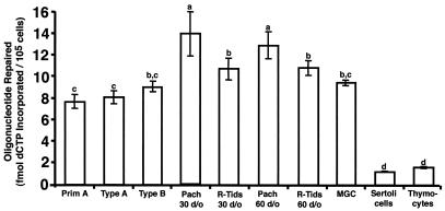
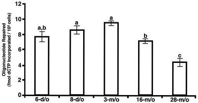
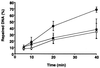
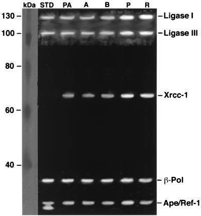
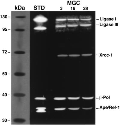
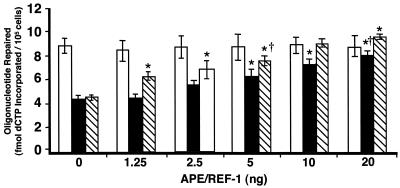
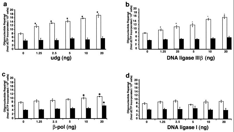
The combined observations of elevated DNA repair gene expression, high uracil-DNA glycosylase-initiated base excision repair, and a low spontaneous mutant frequency for a lacI transgene in spermatogenic cells from young mice suggest that base excision repair activity is high in spermatogenic cell types. Notably, the spontaneous mutant frequency of the lacI transgene is greater in spermatogenic cells obtained from old mice, suggesting that germ line DNA repair activity may decline with age. A paternal age effect in spermatogenic cells is recognized for the human population as well. To determine if male germ cell base excision repair activity changes with age, uracil-DNA glycosylase-initiated base excision repair activity was measured in mixed germ cell (i.e., all spermatogenic cell types in adult testis) nuclear extracts prepared from young, middle-aged, and old mice. Base excision repair activity was also assessed in nuclear extracts from premeiotic, meiotic, and postmeiotic spermatogenic cell types obtained from young mice. Mixed germ cell nuclear extracts exhibited an age-related decrease in base excision repair activity that was restored by addition of apurinic/apyrimidinic (AP) endonuclease. Uracil-DNA glycosylase and DNA ligase were determined to be limiting in mixed germ cell nuclear extracts prepared from young animals. Base excision repair activity was only modestly elevated in pachytene spermatocytes and round spermatids relative to other spermatogenic cells. Thus, germ line short-patch base excision repair activity appears to be relatively constant throughout spermatogenesis in young animals, limited by uracil-DNA glycosylase and DNA ligase in young animals, and limited by AP endonuclease in old animals.
Figures
Similar articles
-
Mutagenesis is elevated in male germ cells obtained from DNA polymerase-beta heterozygous mice.Biol Reprod. 2008 Nov;79(5):824-31. doi: 10.1095/biolreprod.108.069104. Epub 2008 Jul 23. Biol Reprod. 2008. PMID: 18650495 Free PMC article.
-
Highly efficient base excision repair (BER) in human and rat male germ cells.Nucleic Acids Res. 2001 Apr 15;29(8):1781-90. doi: 10.1093/nar/29.8.1781. Nucleic Acids Res. 2001. PMID: 11292851 Free PMC article.
-
Mixed spermatogenic germ cell nuclear extracts exhibit high base excision repair activity.Nucleic Acids Res. 2001 Mar 15;29(6):1366-72. doi: 10.1093/nar/29.6.1366. Nucleic Acids Res. 2001. PMID: 11239003 Free PMC article.
-
Envisioning the molecular choreography of DNA base excision repair.Curr Opin Struct Biol. 1999 Feb;9(1):37-47. doi: 10.1016/s0959-440x(99)80006-2. Curr Opin Struct Biol. 1999. PMID: 10047578 Review.
-
Suppression of spontaneous mutagenesis in human cells by DNA base excision-repair.Mutat Res. 2000 Apr;462(2-3):129-35. doi: 10.1016/s1383-5742(00)00024-7. Mutat Res. 2000. PMID: 10767624 Review.
Cited by
-
Tumor growth is suppressed in mice expressing a truncated XRCC1 protein.Am J Cancer Res. 2012;2(2):168-77. Epub 2012 Feb 15. Am J Cancer Res. 2012. PMID: 22432057 Free PMC article.
-
Stage-specific gene expression is a fundamental characteristic of rat spermatogenic cells and Sertoli cells.Proc Natl Acad Sci U S A. 2008 Jun 17;105(24):8315-20. doi: 10.1073/pnas.0709854105. Epub 2008 Jun 10. Proc Natl Acad Sci U S A. 2008. PMID: 18544648 Free PMC article.
-
Paternal age, de novo mutations, and offspring health? New directions for an ageing problem.Hum Reprod. 2024 Dec 1;39(12):2645-2654. doi: 10.1093/humrep/deae230. Hum Reprod. 2024. PMID: 39361588 Free PMC article. Review.
-
Mutagenesis is elevated in male germ cells obtained from DNA polymerase-beta heterozygous mice.Biol Reprod. 2008 Nov;79(5):824-31. doi: 10.1095/biolreprod.108.069104. Epub 2008 Jul 23. Biol Reprod. 2008. PMID: 18650495 Free PMC article.
-
Non-muscle invasive bladder cancer tissues have increased base excision repair capacity.Sci Rep. 2020 Oct 1;10(1):16371. doi: 10.1038/s41598-020-73370-z. Sci Rep. 2020. PMID: 33004944 Free PMC article.
References
-
- Alcivar, A. A., L. E. Hake, and N. B. Hecht. 1992. DNA polymerase-beta and poly(ADP)ribose polymerase mRNAs are differentially expressed during the development of male germinal cells. Biol. Reprod. 46:201-207. - PubMed
-
- Beard, W. A., and S. H. Wilson. 2000. Structural design of eukaryotic DNA repair polymerase: DNA polymerase β. Mutat. Res. 460:231-244. - PubMed
-
- Bellve, A. R. 1993. Purification, culture, and fractionation of spermatogenic cells. Methods Enzymol. 225:84-113. - PubMed
-
- Bellve, A. R., C. F. Millette, Y. M. Bhatnagar, and D. A. O'Brien. 1977. Dissociation of the mouse testis and characterization of isolated spermatogenic cells. J. Histochem. Cytochem. 25:480-494. - PubMed
Publication types
MeSH terms
Substances
Grants and funding
LinkOut - more resources
Full Text Sources
Medical
Miscellaneous