

Involvement of conserved histidine, lysine and tyrosine residues in the mechanism of DNA cleavage by the caspase-3 activated DNase CAD
- PMID: 11884629
- PMCID: PMC101349
- DOI: 10.1093/nar/30.6.1325
Involvement of conserved histidine, lysine and tyrosine residues in the mechanism of DNA cleavage by the caspase-3 activated DNase CAD
Abstract
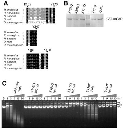
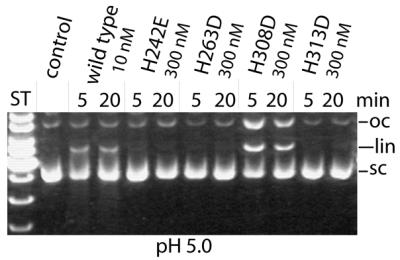
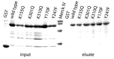
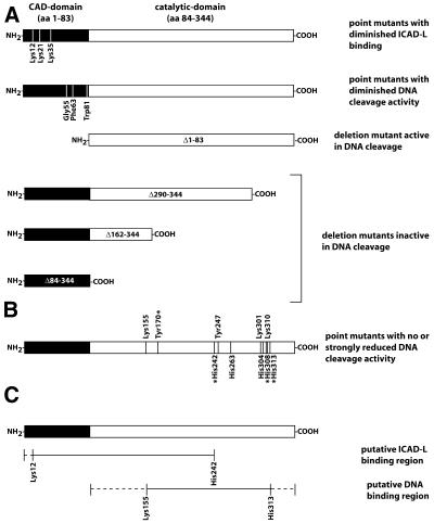
The caspase-activated DNase (CAD) is involved in DNA degradation during apoptosis. Chemical modification of murine CAD with the lysine-specific reagent 2,4,6-trinitrobenzenesulphonic acid and the tyrosine-specific reagent N-acetylimidazole leads to inactivation of the nuclease, indicating that lysine and tyrosine residues are important for DNA cleavage by this enzyme. The presence of DNA or the inhibitor ICAD-L protects the enzyme from modification. Amino acid substitution in murine CAD of lysines and tyrosines conserved in CADs from five different species leads to variants with little if any catalytic activity, but unaltered DNA binding (K155Q, K301Q, K310Q, Y247F), with the exception of Y170F, which retains wild-type activity. Similarly, as observed for the previously characterised H242N, H263N, H308N and H313N variants, the newly introduced His-->Asp/Glu or Arg exchanges lead to variants with <1% of wild-type activity, with two exceptions: H313R shows wild-type activity, and H308D at pH 5.0 exhibits approximately 5% of wild-type activity at this pH. Y170F and H313R produce a specific pattern of fragments, different from wild-type CAD, which degrades DNA non-specifically. The recombinant nuclease variants produced in Escherichia coli were tested for their ability to form nucleolytically active oligomers. They did not show any significant deviation from the wild-type enzyme. Based on these and published data possible roles of the amino acid residues under investigation are discussed.
Figures

Similar articles
-
Identification of functionally relevant histidine residues in the apoptotic nuclease CAD.Nucleic Acids Res. 2001 Oct 1;29(19):3901-9. doi: 10.1093/nar/29.19.3901. Nucleic Acids Res. 2001. PMID: 11574671 Free PMC article.
-
Enzymatic active site of caspase-activated DNase (CAD) and its inhibition by inhibitor of CAD.Arch Biochem Biophys. 2001 Apr 1;388(1):91-9. doi: 10.1006/abbi.2000.2266. Arch Biochem Biophys. 2001. PMID: 11361146
-
Experimental evidence for a beta beta alpha-Me-finger nuclease motif to represent the active site of the caspase-activated DNase.Biochemistry. 2003 Aug 12;42(31):9288-94. doi: 10.1021/bi0348765. Biochemistry. 2003. PMID: 12899615
-
Apoptotic DNA fragmentation.Exp Cell Res. 2000 Apr 10;256(1):12-8. doi: 10.1006/excr.2000.4834. Exp Cell Res. 2000. PMID: 10739646 Review.
-
Degradation of chromosomal DNA during apoptosis.Cell Death Differ. 2003 Jan;10(1):108-16. doi: 10.1038/sj.cdd.4401161. Cell Death Differ. 2003. PMID: 12655299 Review.
Cited by
-
MiRNA-145 Induces Apoptosis in a Gallbladder Carcinoma Cell Line by Targeting DFF45.Open Life Sci. 2018 Jul 5;13:227-235. doi: 10.1515/biol-2018-0027. eCollection 2018 Jan. Open Life Sci. 2018. PMID: 33817087 Free PMC article.
-
Engineered apoptotic nucleases for chromatin research.Nucleic Acids Res. 2007;35(13):e93. doi: 10.1093/nar/gkm486. Epub 2007 Jul 10. Nucleic Acids Res. 2007. PMID: 17626049 Free PMC article.
-
Chromatin collapse during caspase-dependent apoptotic cell death requires DNA fragmentation factor, 40-kDa subunit-/caspase-activated deoxyribonuclease-mediated 3'-OH single-strand DNA breaks.J Biol Chem. 2013 Mar 29;288(13):9200-15. doi: 10.1074/jbc.M112.411371. Epub 2013 Feb 21. J Biol Chem. 2013. PMID: 23430749 Free PMC article.
-
The effect of ICAD-S on the formation and intracellular distribution of a nucleolytically active caspase-activated DNase.Nucleic Acids Res. 2002 Jul 15;30(14):3045-51. doi: 10.1093/nar/gkf431. Nucleic Acids Res. 2002. PMID: 12136086 Free PMC article.
-
The role of the DFF40/CAD endonuclease in genomic stability.Apoptosis. 2021 Feb;26(1-2):9-23. doi: 10.1007/s10495-020-01649-7. Epub 2021 Jan 2. Apoptosis. 2021. PMID: 33387146 Review.
References
-
- Nagata S. (2000) Apoptotic DNA fragmentation. Exp. Cell Res., 256, 12–18. - PubMed
-
- Liu X., Zou,H., Slaughter,C. and Wang,X. (1997) DFF, a heterodimeric protein that functions downstream of caspase-3 to trigger DNA fragmentation during apoptosis. Cell, 89, 175–184. - PubMed
-
- Enari M., Sakahira,H., Yokoyama,H., Okawa,K., Iwamatsu,A. and Nagata,S. (1998) A caspase-activated DNase that degrades DNA during apoptosis, and its inhibitor ICAD. Nature, 391, 43–50. - PubMed
-
- Halenbeck R., MacDonald,H., Roulston,A., Chen,T.T., Conroy,L. and Williams,L.T. (1998) CPAN, a human nuclease regulated by the caspase-sensitive inhibitor DFF45. Curr. Biol., 8, 537–540. - PubMed
-
- Sakahira H., Iwamatsu,A. and Nagata,S. (2000) Specific chaperone-like activity of inhibitor of caspase-activated DNase for caspase-activated DNase. J. Biol. Chem., 275, 8091–8096. - PubMed
Publication types
MeSH terms
Substances
LinkOut - more resources
Full Text Sources
Research Materials
Miscellaneous
