

Heat-induced formation of reactive oxygen species and 8-oxoguanine, a biomarker of damage to DNA
- PMID: 11884633
- PMCID: PMC101361
- DOI: 10.1093/nar/30.6.1354
Heat-induced formation of reactive oxygen species and 8-oxoguanine, a biomarker of damage to DNA
Abstract
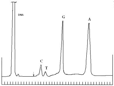
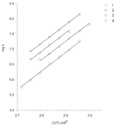
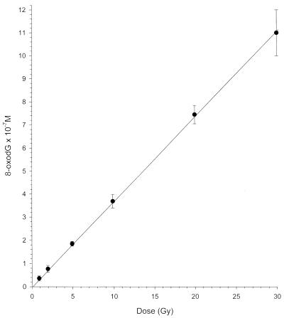
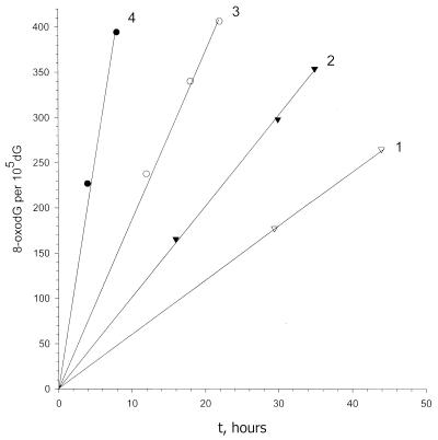
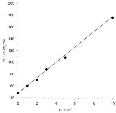
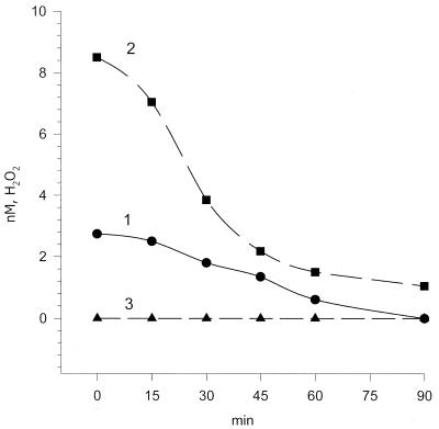
Heat-induced formation of 8-oxoguanine was demonstrated in DNA solutions in 10(-3) M phosphate buffer, pH 6.8, by enzyme-linked immunosorbent assays using monoclonal antibodies against 8-oxoguanine. A radiation-chemical yield of 3.7 x 10(-2) micromol x J(-1) for 8-oxoguanine production in DNA upon gamma-irradiation was used as an adequate standard for quantitation of 8-oxoguanine in whole DNA. The initial yield of heat-induced 8-oxoguanine exhibits first order kinetics. The rate constants for 8-oxoguanine formation were determined at elevated temperatures; the activation energy was found to be 27 +/- 2 kcal/mol. Extrapolation to 37 degrees C gave a value of k37 = 4.7 x 10(-10) x s(-1). Heat-induced 8-oxoguanine formation and depurination of guanine and adenine show similarities of the processes, which implies that heat-mediated generation of reactive oxygen species (ROS) should occur. Heat-induced production of H2O2 in phosphate buffer was shown. The sequence of reactions of thermally mediated ROS formation have been established: activation of dissolved oxygen to the singlet state, generation of superoxide radicals and their dismutation to H2O2. Gas saturation (O2, N2 and Ar), D2O, scavengers of 1O2, O2-* and OH* radicals and metal chelators influenced heat-induced 8-oxoguanine formation as they affected thermal ROS generation. These findings imply that heat acts via ROS attack leading to oxidative damage to DNA.
Figures
References
-
- Beckman K.B. and Ames,B.N. (1998) The free radical theory of aging matures. Physiol. Rev., 78, 547–581. - PubMed
-
- Kuchino Y., Mori,F., Kasai,H., Inone,H., Iwai,S., Miure,K., Ohtsuka,E. and Nishimura,S. (1987) Misreading of DNA templates containing 8-hydroxydeoxyguanosine at the modified base and at adjacent residues. Nature, 327, 77–79. - PubMed
-
- Shibutani S., Takeshita,M. and Grollman,A.P. (1991) Insertion of specific bases during DNA synthesis past the oxidation-damaged base 8-oxoG. Nature, 349, 431–434. - PubMed
-
- Pavlov Y.I., Minnick,D.T., Izuta,S. and Kunkel,T.A. (1994) DNA replication fidelity with 8-oxodeoxyguanosine triphosphate. Biochemistry, 33, 4695–4701. - PubMed
-
- Bruskov V.I. and Kuklina,O.V. (1988) 8-Oxo-GTP displays substrate properties of UTP in polynucleotide synthesis catalyzed by Escherichia coli RNA polymerase on a poly[d(A-T)]·poly[d(A-T)] template. Mol. Biol., 22, 580–584. - PubMed
Publication types
MeSH terms
Substances
LinkOut - more resources
Full Text Sources
Other Literature Sources
Research Materials
