Comparative Study
. 2002 Feb;14(2):421-33.
doi: 10.1105/tpc.010282.

AXR1-ECR1-dependent conjugation of RUB1 to the Arabidopsis Cullin AtCUL1 is required for auxin response

Affiliations
Comparative Study

AXR1-ECR1-dependent conjugation of RUB1 to the Arabidopsis Cullin AtCUL1 is required for auxin response

Juan C del Pozo et al. Plant Cell. 2002 Feb.

Abstract

Mutations in the AXR1 gene result in a reduction in auxin response and diverse defects in auxin-regulated growth and development. In a previous study, we showed that AXR1 forms a heterodimer with the ECR1 protein. This enzyme activates the ubiquitin-related protein RUB1 in vitro. Furthermore, we showed that the Skp1-Cul1/Cdc53-F-box (SCF) subunit AtCUL1 is modified by RUB1 in vivo. In this report, we demonstrate that the formation of RUB-AtCUL1 is dependent on AXR1 and ECR1 in vivo. The expression of AXR1 and ECR1 is restricted to zones of active cell division and cell elongation, consistent with their role in growth regulation. These results provide strong support for a model in which RUB conjugation of AtCUL1 affects the function of SCF E3s that are required for auxin response.


Figures

Figure 1.
Figure 1.
The AXR1 Gene Is Expressed in Dividing and Elongating Cells throughout the Plant. Expression was determined by in situ localization of RNA ([A] to [D], [H], and [I]) and immunolocalization of the AXR1 protein ([E] to [G] and [J] to [P]). (A) Root meristem hybridized with antisense AXR1 RNA. (B) Root meristem hybridized with sense AXR1 RNA. (C) Seven-day-old seedling hybridized with antisense AXR1 RNA. Staining is visible on the leaf primordium. (D) Seven-day-old seedling hybridized with sense AXR1 RNA. (E) Section of a shoot meristem stained with antibodies against AXR1. (F) Section of an axr1-12 shoot meristem stained with antibodies against AXR1. (G) Cross-section of the root elongation zone stained with antibodies against AXR1. The arrow indicates a stained nucleus in the epidermis. (H) Inflorescence and floral meristems hybridized with antisense AXR1 RNA. (I) Inflorescence and floral meristems hybridized with sense AXR1 RNA. (J) Inflorescence meristem (im) and floral meristems stained with AXR1 antibodies. II and VII indicate stages of flower development. (K) Higher magnification of a stage II flower stained with AXR1 antibodies. (L) Inflorescence meristem and flower section of axr1-12 stained with AXR1 antibodies. (M) Wild-type zygote cell stained with AXR1 antiserum. (N) Proembryo stage stained with AXR1 antibodies. The brown color in the endothecium (Eth) is not the result of AXR1 staining. (O) Forty-hour-old embryo stained with AXR1 antibodies. (P) Early globular embryo stained with AXR1 antibodies. Stained nuclei in the suspensor cell are detected. Bars in (A) to (L) = 50 μm.
Figure 2.
Figure 2.
Pattern of AXR1 Expression as Revealed by Staining AXR1::GUS Seedlings. (A) GUS staining of a 7-day-old light-grown seedling. (B) GUS staining of a 7-day-old light-grown root. (C) Three-day-old dark-grown seedling. Note the higher expression of AXR1 on the lower side of the apical hook than on the upper zone. (D) Developing flowers. (E) A 0.5-day-old seedling. The bottom panel shows the same seedling after mechanically removing the seed coat. (F) Primary leaf of a 7-day-old light-grown seedling stained for GUS activity. Note that AXR1 is highly expressed in the young developing trichomes. (G) Root hairs at the beginning of the differentiation zone of a 7-day-old seedling. (H) Mature leaf (20 days old) of a light-grown plant stained for GUS activity. The arrows indicate the hydathodes.
Figure 3.
Figure 3.
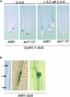
AXR1 Plays an Important Role in Lateral Root Development. (A) AXR1 is required for the expression of CycB1;1 during lateral root formation. Five-day-old AXR1 CycB1;1::GUS, and axr1-12 CycB1;1::GUS seedlings were grown in the absence (−) or presence (+) of 0.2 μM 2,4-D for 24 hr and stained for GUS activity. (B) Left, GUS expression in lateral root primordia formed after treatment with 0.01 μM 2.4-D for 24 hr. Arrows indicate the positions of lateral root primordia. Center, GUS staining in pericycle cells before cell division in a 2,4-D–treated root. Right, GUS staining in dividing cells in a later stage of lateral root development and in the vascular tissue close to the primordium.
Figure 4.
Figure 4.
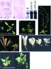
ECR1 Functions with AXR1 during Plant Development. (A) Antisense ECR1 probe hybridized to a section of inflorescence and developing flowers. (B) Sense probe hybridized to a similar section. (C) RNA gel blot showing ECR1 expression in 7-day-old wild-type (wt) seedlings and transgenic lines expressing ECR1C215A. (D) Rosettes of ECR1-C4 and wild-type plants at 21 days. (E) Phenotypes of ECR1C215A transgenic plants at 42 days. (F) Phenotype of a wild-type plant at 42 days. (G) Inflorescence with mature siliques of ECR1C215A transgenic plants. (H) Flowers of wild type (i), axr1-12 (ii), ECR1-C1 (iii), and the axr1-12 ECR1-C1 double mutant (iv). (I) Rosettes of axr1-12 ECR1-C1 double mutant plants at 42 days. Bars in (D) to (F), and (I) = 1 cm.
Figure 5.
Figure 5.
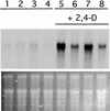
Auxin-Induced Gene Expression Is Reduced in ECR1C215A Transgenic Plants. RNA gel blot analysis of total RNA extracted from wild-type (lanes 1 and 5), ECR1-C4 (lanes 2 and 6), ECR1-C1 (lanes 3 and 7), and axr1-12 (lanes 4 and 8) plants treated with or without 20 mM 2,4-D. The blot was hybridized to IAA2 probe. An ethidium bromide–stained gel is shown at bottom.
Figure 6.
Figure 6.
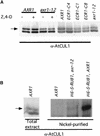
AXR1 and ECR1 Are Required for Modification of AtCUL1. (A) Protein gel blot analysis of total proteins extracted from wild-type, axr1-12, and ECR1C215A transgenic plants. Extracts were prepared from 6-day-old seedlings, and either 10 mg (first four lanes) or 5 mg (next five lanes) was loaded onto the gel. α-AtCUL1, AtCUL1 antiserum. (B) Protein gel blot analysis of total extract or nickel-purified proteins recovered from AXR1 and axr1-12 5-day-old seedlings carrying the H6-S–RUB1 transgene. The blots were probed with AtCUL1 antiserum (α-AtCUL1). The arrow indicates the position of RUB-AtCUL1.
Figure 7.
Figure 7.
The axr1 Mutation Does Not Affect the Nuclear Localization of AtCUL1. Immunolocalization of AtCUL1 in wild-type (A) and axr1-12 (B) floral sections. The insets show high magnification images from the carpel walls, demonstrating normal nuclear localization in the mutant. The AtCUL1 antiserum was characterized previously (Gray et al., 1999; Farras et al., 2001).

Similar articles

Cited by

References

    1. Abel, S., and Theologis, A. (1996). Early genes and auxin action. Plant Physiol. 111, 9–17. - PMC - PubMed
    1. Abel, S., Nguyen, M.D., and Theologis, A. (1995). The PS-IAA4/ 5-like family of early auxin-inducible mRNAs in Arabidopsis thaliana. J. Mol. Biol. 251, 533–549. - PubMed
    1. Ausubel, F.M., Brent, R., Kingston, R.E., Moore, D.D., Seidman, J.G., Smith, J.A., and Struhl, K. (1990). Current Protocols in Molecular Biology. (New York: Wiley).
    1. Bechtold, N., Ellis, J., and Pelletier, G. (1993). In planta Agrobacterium mediated gene transfer by infiltration of adult Arabidopsis thaliana plants. C. R. Acad. Sci. Paris Life Sci. 316, 15–18.
    1. Berleth, T., Mattsson, J., and Hardtke, C.S. (2000). Vascular continuity and auxin signals. Trends Plant Sci. 5, 387–393. - PubMed

Publication types

MeSH terms