. 2002 Mar 15;21(6):1255-66.
doi: 10.1093/emboj/21.6.1255.

Conserved segments 1A and 2B of the intermediate filament dimer: their atomic structures and role in filament assembly

Affiliations

Conserved segments 1A and 2B of the intermediate filament dimer: their atomic structures and role in filament assembly

Sergei V Strelkov et al. EMBO J. .

Abstract

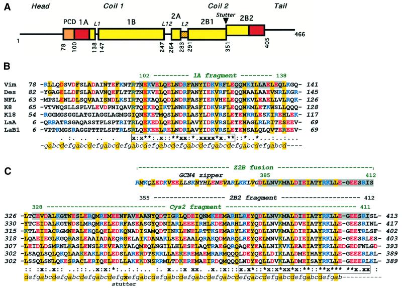
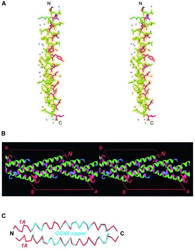
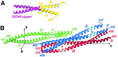
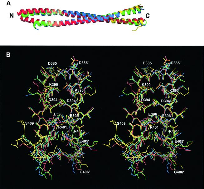
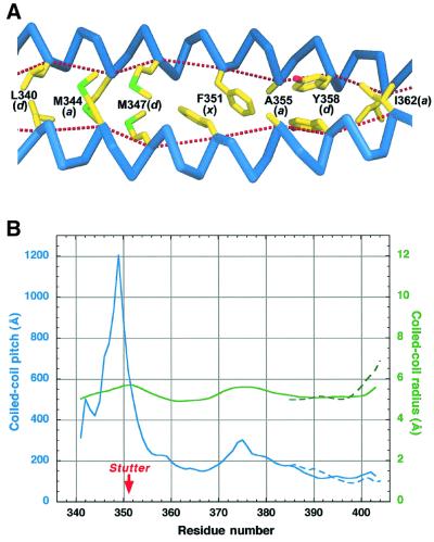
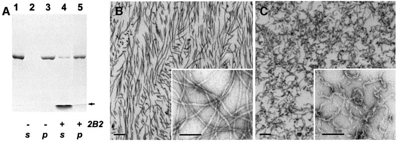
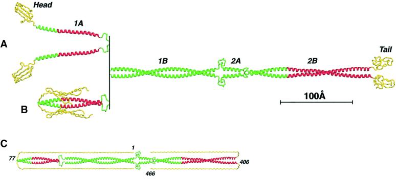
Intermediate filaments (IFs) are key components of the cytoskeleton in higher eukaryotic cells. The elementary IF 'building block' is an elongated coiled-coil dimer consisting of four consecutive alpha-helical segments. The segments 1A and 2B include highly conserved sequences and are critically involved in IF assembly. Based on the crystal structures of three human vimentin fragments at 1.4-2.3 A resolution (PDB entries 1gk4, 1gk6 and 1gk7), we have established the molecular organization of these two segments. The fragment corresponding to segment 1A forms a single, amphipatic alpha-helix, which is compatible with a coiled-coil geometry. While this segment might yield a coiled coil within an isolated dimer, monomeric 1A helices are likely to play a role in specific dimer-dimer interactions during IF assembly. The 2B segment reveals a double-stranded coiled coil, which unwinds near residue Phe351 to accommodate a 'stutter'. A fragment containing the last seven heptads of 2B interferes heavily with IF assembly and also transforms mature vimentin filaments into a new kind of structure. These results provide the first insight into the architecture and functioning of IFs at the atomic level.


Figures

None
Fig. 1. (A) Primary structure of IF proteins. Schematic diagram of human vimentin. Rectangles show α-helical segments, including the pre-coil domain (PCD). (B) Sequence alignment of the 1A segments of human IF proteins including vimentin, desmin, neurofilament L protein, cytokeratins 8 and 18, and nuclear lamins A and B1. (C) Similar alignment of the 2B segments. Vimentin fragments 1A, Cys2, Z2B and 2B2 are highlighted. The heptad repeats are marked as abcdefg, with core positions highlighted with yellow. Basic and acidic residues are shown in blue and red, respectively. The line below the alignment shows the sequence similarity score s of a particular residue in the seven proteins: ‘*’, s = 1.0 (absolutely conserved); ‘x’, 0.75≤s<1.0; ‘:’, 0.5≤s<0.75; ‘.’, 0.25≤s<0.5 (see Materials and methods for details). The two most conserved regions within the 1A segment and in the C-terminal part of the 2B segment, respectively, are shown in boxes.
None
Fig. 2. Crystal structure of the vimentin fragment 1A. (A) Stereo view of the atomic model and electron density map with coefficients 2FobsFcalc contoured at 1.2σ. Residues in the a and d positions of the putative heptad repeat are shown in magenta. Solvent molecules are shown as blue spheres. (B) The crystal packing arrangement of 1A shown in stereo. The a and d positions are highlighted with magenta. The N- and C-termini of the helices are marked in red and blue, respectively. (C) Modeling of a parallel coiled-coil by docking two 1A helices (red) while using the GCN4 zipper structure (cyan) as a ruler.
None
Fig. 3. Crystal structures of the Z2B and Cys2 fragments. (A) Ribbon diagram of the Z2B structure. The GCN4 leucine zipper and the authentic vimentin residues are shown in magenta and yellow, respectively. (B) Ribbon diagrams of the three symmetry-independent Cys2 dimers AB (red), CD (green) and EF (blue) in the unit cell. The position of the stutter in each dimer is marked with an asterisk.
None
Fig. 4. Superposition of the Cys2 and Z2B structures. (A) Ribbon diagrams of the three Cys2 dimers (AB, red; CD, green; and EF, blue) and the Z2B dimer (yellow). (B) The C-terminal part of the 2B segment shown in stereo (coloring as above). Salt bridges are shown with dotted lines.
None
Fig. 5. Effect of the coiled-coil stutter within the 2B segment. (A) Hydrophobic core organization near the stutter of the Cys2 dimer. The letters in parentheses after the residue number indicate the heptad position. The dotted lines connect the Cα atoms of the consecutive core residues. (B) Coiled-coil radius (green) and pitch (blue) as a function of residue number. The data for the Cys2 structure (averages over three independent dimers) and Z2B structure are shown with solid and dashed lines, respectively.
None
Fig. 6. Effect of the 2B2 fragment on IF assembly. (A) Denaturing gel electrophoresis (SDS–PAGE) illustrating the assembly of human recombinant vimentin alone (lanes 2 and 3) and in the presence of the 2B2 fragment (lanes 4 and 5). The 2B2 fragment was added to the vimentin sample (lane 1) at a 10-fold molar excess, and then filament assembly in the test and reference (i.e. without the fragment addition) samples was performed as described in Materials and methods. The samples subsequently were centrifuged in a Beckman Airfuge for 30 min at 10 p.s.i. yielding the supernatant (lanes 2 and 4) and pelleted (lanes 3 and 5) fractions. The arrow indicates the location of the gel front. (B) Negatively stained EM images of vimentin IFs assembled in vitro. The samples were prepared by ultrathin sectioning (main figure) or on grids (inset). Scale bars are 100 nm. (C) Similarly assembled IFs, which subsequently were incubated with a 10-fold molar excess of the 2B2 fragment for 1 h at 37°C.
None
Fig. 7. Atomic model of the IF dimer. (A) ‘Open’ conformation of the 1A segments. Regions corresponding to the three described crystal structures are shown in red. (B) ‘Closed’ conformation of the 1A segments. (C) Modeling of the fully extended conformations of the head and tail domains (yellow).

References

    1. Aebi U., Haner,M., Troncoso,J., Eichner,R. and Engel,A. (1988) Unifying principles in intermediate filament structure and assembly. Protoplasma, 145, 73–81.
    1. Brown J.H., Cohen,C. and Parry,D.A. (1996) Heptad breaks in α-helical coiled coils: stutters and stammers. Proteins, 26, 134–145. - PubMed
    1. Brünger A.T. and Nilges,M. (1993) Computational challenges for macromolecular structure determination by X-ray crystallography and solution NMR-spectroscopy. Q. Rev. Biophys., 26, 49–125. - PubMed
    1. Brünger A.T. et al. (1998) Crystallography and NMR system: a new software suite for macromolecular structure determination. Acta Crystallogr. D, 54, 905–921. - PubMed
    1. Burkhard P., Kammerer,R.A., Steinmetz,M.O., Bourenkov,G.P. and Aebi,U. (2000) The coiled-coil trigger site of the rod domain of cortexillin I unveils a distinct network of inter- and intra-helical salt-bridges. Structure, 8, 223–230. - PubMed

Publication types